 by
by Life begins within cells, where different organelles play vital roles in maintaining optimal cell functionality. Two such essential organelles are Golgi bodies and mitochondria. These structures are distinct in their functions and structures but share a vital role in ensuring cellular health and vitality.
Definition of Golgi Bodies and Mitochondria?
Sure! Here are the definitions of Golgi Bodies and Mitochondria:
Golgi Bodies (Golgi Apparatus): Golgi Apparatus or Complex can be found within most eukaryotic cells and tissues. They play a vital role in cell divisions and division of metabolic pathways. The endomembrane systems, which include the Golgi apparatus (ER), lysosomes, and vesicles, are essential to keeping our bodies at their maximum capacity. The Golgi Bodies play a crucial role in processing, modifying, and packaging proteins and lipids before they are transported to their final destinations within or outside the cell.
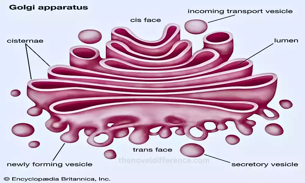
Structure: The Golgi Bodies consist of a series of flattened, membrane-bound sacs called cisternae. These cisternae are stacked on each other, forming Golgi stacks or dictyosomes. The Golgi apparatus may have several stacks in a cell, depending on its function and cell type.
Function: The primary functions of the Golgi Bodies include modifying and sorting proteins and lipids synthesized in the endoplasmic reticulum. Further modifying these molecules through various posttranslational modifications such as glycosylation (adding sugar groups) and phosphorylation (adding phosphate groups).
After modification, the proteins and lipids are sorted into specific vesicles for transport to their intended destinations, such as the plasma membrane, lysosomes, or secretion outside the cell.
Mitochondria: Mitochondria are organelles that can be found in most eukaryotic (excluding red blood cells) cells. They have double membranes with rod-shaped shapes and are found inside eukaryotic cellular structures.
Their primary responsibility lies in producing energy via Adenosine Triphosphate (ATP), the currency for cell metabolism through respiratory carbonic anhydrase activity.
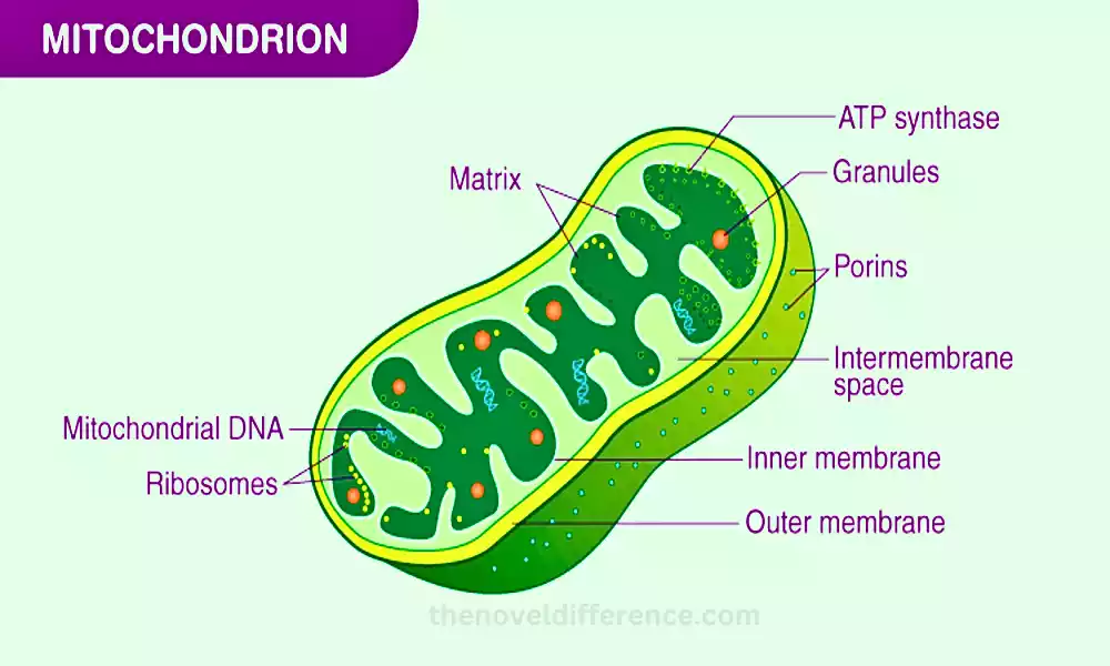
Structure: Mitochondria have an outer membrane and an inner membrane, which contains numerous infoldings called cristae. The space enclosed by the inner membrane is known as the mitochondrial matrix. Mitochondria have their own DNA and ribosomes for protein production.
Function: Mitochondria’s primary role is producing ATP through aerobic respiration – breaking down glucose and other molecules with oxygen to produce energy for our cells to use as fuel. This process takes place in the inner mitochondrial membrane and the mitochondrial matrix. ATP generated by mitochondria is used to power various cellular processes, including muscle contractions, active transport, and biosynthesis.
At a cellular level, mitochondria play an integral part in numerous other ways as well. From controlling calcium levels and playing their part in programmed cell death via apoptosis while producing reactive oxygen species (ROS).
Golgi Bodies and Mitochondria both play vital roles in cell function; with Golgi Bodies focused on protein processing and transport and Mitochondria taking responsibility for producing energy through ATP production and energy metabolism respectively.
What are Golgi Bodies?
Golgi Bodies also referred to as Golgi Apparatus or Complex, are organelles found within eukaryotic cells and comprise their Golgi Apparatus or Complex. The endomembrane systems, which include the Golgi apparatus (ER), lysosomes, and vesicles, are dominated by lysosomes.
Golgi Bodies play a crucial role in processing, modifying, and packaging proteins and lipids before they are transported to their final destinations within or outside the cell.
1. Structure:
The Golgi Bodies consist of a series of flattened, membrane-bound sacs called cisternae. These cisternae are stacked on each other, forming Golgi stacks or dictyosomes. The Golgi apparatus may have several stacks in a cell, depending on its function and cell type.
Each Golgi stack is composed of several compartments with distinct functions, including the cis-Golgi network, medial cisternae, and trans-Golgi network. Vesicles shuttle between these compartments, transporting molecules for further processing and sorting.
2. Function:
The primary functions of Golgi Bodies include:
Protein Modification: Golgi Bodies receive proteins from the endoplasmic reticulum (ER) in vesicles. Once inside the Golgi, these proteins undergo various post-translational modifications. These modifications can include glycosylation, phosphorylation, or sulfation. These modifications are crucial for the maturation of protein function.
Protein Sorting: Golgi Bodies sort and direct proteins to their appropriate cellular destinations. Some proteins are targeted for secretion outside the cell, and Golgi facilitates their packaging into secretory vesicles that eventually merge with the plasma membrane for exocytosis.
Lipid Processing: Apart from processing proteins, Golgi Bodies also modify and sort lipids, which are essential components of cell membranes and play various signaling roles within the cell.
Formation of Lysosomes and Secretory Vesicles: Golgi Bodies are involved in the formation of lysosomes, which are cellular organelles responsible for intracellular digestion. They package hydrolytic enzymes into vesicles called lysosomes. Additionally, Golgi participates in the formation of secretory vesicles that carry newly processed and sorted molecules to the cell surface for secretion.
Golgi Bodies are crucial organelles that play a central role in the post-translational modification, sorting, and packaging of proteins and lipids, ensuring their proper distribution and functioning within the cell and for secretion outside the cell. They are essential for maintaining the cell’s overall functionality and homeostasis.
Structure of Golgi Bodies
Golgi Bodies (commonly referred to as Golgi Apparatus or Complex) consist of flattened membrane sacs called cisternae that connect each cell in its structure. These cisternae are interlinked to form an intricate network within a cell’s infrastructure, creating an organized yet complex structure within that cell.
Here is a more detailed overview of the structure of Golgi Bodies:
1. Cisternae:
Golgi Bodies consist of several individual cisternae, which are flattened, curved membrane sacs. These cisternae are arranged in parallel stacks, and each stack may contain multiple cisternae. The Golgi stacks can vary in number depending on the specific cell type and its functional requirements.
2. Golgi Stacks:
Golgi stacks (commonly referred to as dictyosomes) can vary from several dozen in number in a eukaryotic cell and can often be found close to its nucleus; their convex face often points towards the endoplasmic reticulum (ER), with their concave side facing towards the plasma membrane.
3. Cisternal Maturation Model:
The cisternal maturation model describes the dynamic nature of the Golgi stacks. It suggests that the cisternae of the Golgi apparatus are not fixed structures but rather are constantly forming and maturing.
New cisternae are continuously added at the cis-Golgi network (CGN) or cis-Golgi, while older cisternae progress through the stack, eventually maturing into the trans-Golgi network (TGN) or trans-Golgi.
4. Golgi Compartments:
The Golgi apparatus is functionally divided into three regions or compartments:
a. Cis-Golgi Network (CGN): The CGN is located near the endoplasmic reticulum and acts as the entry point for proteins and lipids arriving from the ER in transport vesicles. It is involved in the initial processing of these molecules.
b. Medial Cisternae: These are the middle compartments of the Golgi stack. The proteins and lipids that arrive at the CGN are modified and processed as they move through the medial cisternae.
c. Trans-Golgi Network (TGN): The TGN is located at the opposite end of the Golgi stack, facing the plasma membrane. It is involved in the final processing and sorting of molecules, preparing them for their specific destinations within or outside the cell.
5. Vesicles and Tubules:
Throughout the Golgi apparatus, there are numerous small transport vesicles and tubules. These vesicles shuttle molecules between the different compartments of the Golgi and are responsible for carrying the processed and sorted proteins and lipids to their appropriate destinations.
The Golgi Bodies have a complex and dynamic structure consisting of stacked cisternae, which are functionally divided into distinct compartments. The Golgi apparatus plays a vital role in processing, modifying, and sorting proteins and lipids, ensuring their proper distribution within the cell and for secretion outside the cell.
Functions of Golgi Bodies
Golgi Bodies, also referred to as Golgi Apparatus or Complex, serve several key roles within eukaryotic cells. These functions primarily involve the processing, modification, sorting, and packaging of proteins and lipids before they are transported to their final destinations within or outside the cell.
The Golgi Bodies play a central role in the endomembrane system and are crucial for maintaining cellular homeostasis and functionality.
Here are the main functions of Golgi Bodies:
1. Protein Modification: Golgi Bodies serve a key function by modifying proteins synthesized by endoplasmic reticulum cells (ERs). These modifications are essential for the functional maturation of proteins. Common modifications include glycosylation, phosphorylation, and sulfation. Glycosylation involves the addition of sugar groups to proteins, which can affect their stability, function, and targeting.
2. Protein Sorting: Golgi Bodies are involved in sorting and directing proteins to their appropriate cellular destinations. Proteins that enter the Golgi apparatus from the ER are further processed and tagged with specific molecular markers, allowing them to be sorted into specific transport vesicles destined for different cellular locations. Some proteins are targeted for secretion outside the cell while others are directed toward organelles such as lysosomes within cells.
3. Lipid Processing: In addition to processing proteins, Golgi Bodies also modify and sort lipids, which are crucial components of cell membranes and play various signaling roles within the cell. The Golgi apparatus is involved in the synthesis of certain lipids and modifies others before their incorporation into cell membranes.
4. Formation of Lysosomes and Secretory Vesicles: Golgi Bodies are responsible for the formation of lysosomes, which are cellular organelles containing hydrolytic enzymes for intracellular digestion. Golgi stacks package these enzymes into vesicles, which then mature into functional lysosomes. Similarly, Golgi Bodies participate in the formation of secretory vesicles, which carry newly processed and sorted molecules to the cell surface for secretion.
5. Vesicle Trafficking and Transport: The Golgi apparatus acts as a central hub for vesicle trafficking and transport within the cell. Vesicles shuttle between the different compartments of the Golgi, transporting molecules for further processing and sorting. This vesicular transport is essential for the efficient movement of proteins and lipids through the endomembrane system.
6. Glycoproteins & Glycolipids Formation: Golgi bodies are responsible for adding carbohydrate molecules (glycoproteins and glycolipids) via glycosylation to proteins & lipids to produce glycosylated molecules which play multiple functions within cells such as cell signaling, immune responses, and cell recognition as well as recognition by other cells.
In summary, the Golgi Bodies are multifunctional organelles involved in the post-translational modification, sorting, and packaging of proteins and lipids. They ensure that these molecules are properly processed and directed to their specific cellular destinations, contributing to various cellular processes essential for cell survival and function.
What are Mitochondria?
Mitochondria are rod-shaped organelles with two membranes and are found within all eukaryotic cells (cells that possess nuclei). Mitochondria are classified as membraned organelles since their shape resembles two rods connected together with membranes extending out in both directions forming two membrane-bound compartments.
Organelles such as microcytes are found within both eukaryotic (cells with nuclei), and other non-eukaryotic cells with membrane-bound organelles.
The mitochondrial features include:
1. Double Membrane structure: Mitochondria are made up of two membranes, an outer and inner membrane. The outer membrane acts as a barrier while the inner membrane forms cristae structures. The cristae increase the available surface area for chemical reactions that are involved in energy production.
2. Matrix: The mitochondrial matrix is the space enclosed by the inner mitochondrial membrane. The matrix contains enzymes as well as other molecules that are necessary for various metabolic pathways. These include the citric cycle (also called the Krebs cycle) or the oxidation of fatty acids.
3. DNA & Ribosomes: The mitochondria have their own DNA called mitochondrial DNA and ribosomes. Mitochondrial mtDNA is required to produce some of the essential proteins for the organelle’s function. The presence of mitochondrial ribosomes and DNA proves the endosymbiotic theory, which states that mitochondria originated as bacteria that were swallowed up into ancestral eukaryotic cell colonies during evolution in a mutualistic partnership relationship.
Mitochondrial Functions:
4. Cellular respiration: Mitochondria’s primary function is the conversion of nutrients such as glucose and fatty acid into ATP – the currency used by cells. The mitochondrial matrix or inner membrane serves as the center for cellular respiration, an activity comprised of glycolysis and citric acid cycles. This process provides the energy needed for different cellular functions, including muscle contractions and active transport.
5. Mitochondria’s Role in Apoptosis: Mitochondria are essential players in the apoptosis – or programmed cell death – process. When cells become injured beyond repair or undergo normal development processes, their mitochondria release molecules which activate an event cascade leading to cell death and lead to its eventual destruction. This process is crucial in removing unwanted or damaged cells and helps maintain tissue homeostasis.
6. Calcium Regulating: Mitochondria play an important role in the regulation of calcium ions inside cells. They can release and take up calcium ions as a response to cell signals. This helps control calcium concentration and its role in many cellular processes including muscle contractions and cell signaling.
7. Summary: Mitochondria are essential in producing energy through respiration for eukaryotic cells. They are vital for cell survival and function because of their unique double membrane structure, DNA, ribosomes, and other cellular processes.
Structure of Mitochondria
The structure of mitochondria is characterized by a double-membraned organelle with a specific organization that enables its essential functions, particularly energy production through cellular respiration. Let’s explore the key structural components of mitochondria:
1. Outer Membrane:
The mitochondrion has an outer membrane that surrounds the entire organelle. This membrane is porous and contains special transport proteins called porins that allow ions and small molecules to pass into the intermembrane space through pores in it.
2. Intermembrane Space:
The space between the outer and inner membranes is known as the intermembrane space. This region plays a role in the transport of molecules and ions into the inner membrane and helps establish the proton gradient required for ATP synthesis.
3. Inner Membrane:
The inner membrane of the mitochondrion is highly convoluted and folded, forming structures called cristae. These cristae increase the surface area of the inner membrane, providing a vast area for cellular respiration reactions to take place. The inner membrane is impermeable to most ions and molecules and houses many protein complexes involved in the electron transport chain, a critical step in ATP production.
4. Cristae:
Cristae are the infoldings of the inner mitochondrial membrane. They come in various shapes and sizes and create a series of compartments within the mitochondrion. The presence of cristae significantly enhances the efficiency of ATP production by increasing the surface area for the assembly of respiratory enzyme complexes.
5. Matrix:
The space enclosed by the inner mitochondrial membrane is called the mitochondrial matrix. The matrix is a gel-like substance containing enzymes that are essential for various metabolic reactions, including the citric acid cycle (Krebs cycle) that generates energy from glucose and fatty acids. Additionally, mitochondria contain their own DNA (mtDNA), ribosomes, and transfer RNA molecules which play key roles in protein synthesis of certain mitochondrial proteins.
5. Mitochondrial DNA (mtDNA):
Mitochondria contain their own form of DNA called mitochondrial DNA which differs substantially from what’s present within cells’ nuclei. Mitochondrial DNA is circular, and it contains genes that encode some essential proteins involved in the organelle’s energy production process. This self-replicating DNA plays a crucial role in maintaining mitochondrial function and is inherited only from the mother during reproduction.
6. Ribosomes:
The mitochondria contain smaller ribosomes than those present in the cell’s cytoplasm; these synthesize proteins encoded by mitochondrial DNA.
The structure of mitochondria is characterized by a double-membraned organelle with an outer membrane, an inner membrane with cristae, and a matrix containing enzymes, mtDNA, and ribosomes.
Mitochondria are essential organelles that use their unique structure to facilitate efficient cell respiration, producing energy as ATP for cells’ energy requirements and supporting other essential cellular processes.
Functions of Mitochondria
Mitochondria play a multifunctional role in cellular processes. They are primarily responsible for generating ATP, the main currency of the cells through cellular respiration. mitochondria play an essential role in many vital bodily processes and activities; here are a few major mitochondrial functions.
1. ATP production (Cellular respiration): Cellular respiration is the most fundamental and well-known function of mitochondria. The breakdown of nutrients such as glucose and fat acids in the presence of oxygen releases energy. The chemical reactions take place in the inner membrane of the mitochondria, more specifically, within the cristae and mitochondrial matrix. ATP is produced through oxidative phosphorylation. This process involves the electron transportation chain and chemiosmotic couple.
2. Calcium Regulation: Mitochondria play a crucial role in regulating intracellular calcium levels. They can act as calcium storage and release calcium ions rapidly in response to cell signals. Calcium regulation is important for maintaining cell homeostasis. It is also involved in many cellular processes including muscle contractions and cell signaling.
3. Apoptosis: Mitochondria are key in programmed cell death or apoptosis. When cells undergo this form of programmed death, mitochondria release molecules like Cytochrome C into their respective cytoplasm. This causes a series of events that lead to cell death. Cell death is necessary for normal development, tissue balance, and elimination of unwanted or damaged cells.
4. Production of Reactive Oxygen species (ROS): Superoxide radicals and reactive oxygen species (ROS) are produced when incomplete reduction of some oxygen molecules during cell respiration occurs, creating superoxide radicals and reactive oxygen species which while potentially toxic at higher concentrations also signal various cellular processes, including immune responses or cell growth. Although such ROS levels can damage cells at times, their role as messenger molecules plays an important part in signaling various aspects of health such as immune responses.
5. Fatty acid oxidation: Mitochondria participate in the oxidation and breakdown of fatty acids to produce energy. This process is known as beta-oxidation and occurs in the mitochondrial matrix. It is especially important during prolonged fasting periods or intense exercise when glucose supplies are low.
6. Heme Synthesis: Mitochondria play a role in the synthesis and transport of oxygen in red blood cells. They also synthesize other proteins that are involved in electron transport, cellular respiration, and heme.
7. Steroid Synthesis: The adrenal glands contain mitochondria that produce certain steroid hormones such as cortisol and aldosterone.
Mitochondria are organelles that perform multiple crucial roles within our bodies and have numerous essential responsibilities. They provide the cells with energy through their primary role of ATP production via cellular respiration. In addition, mitochondria play a role in calcium regulation and ROS production. They also participate in apoptosis and fatty acid oxidation.
Differences between Golgi Bodies and Mitochondria
Golgi Bodies are essential organelles in eukaryotic cells; however, their structure, function, and location vary significantly among species.
Here are the key differences between Golgi Bodies and Mitochondria:
1. Structure:
Golgi Bodies: The Golgi Body consists of flattened membrane-bound sacs called cisternae that are stacked one on top of another in the juxtanuclear region of cells.
They are part of the endomembrane system, with distinct cis-Golgi and trans-Golgi faces. Golgi Bodies have distinct compartments: cis-Golgi network, medial cisternae, and trans-Golgi network. Vesicles shuttle between these compartments, transporting molecules for further processing and sorting.
Mitochondria: Mitochondria are double membrane structures consisting of an outer and an inner membrane with many infoldings known as c cristae, all enclosed within their own double envelopes. They are typically scattered throughout the cytoplasm of the cell. The space enclosed by the inner mitochondrial membrane is known as the mitochondrial matrix, containing enzymes involved in various metabolic reactions.
2. Function:
Golgi Bodies: Golgi Bodies are involved in processing, modifying, and sorting proteins and lipids. Posttranslational modifications such as glycosylation and phosphorylation take place here. Golgi Bodies then sort the proteins into vesicles for transport to specific cellular destinations, such as the plasma membrane or lysosomes.
Mitochondria: Mitochondria are essential energy producers within cells through cellular respiration, producing ATP from glucose and fatty acids to generate power for energy use in cell functions such as brain activity. They are often referred to as the “powerhouses” of the cell because ATP is the primary energy currency for cellular processes. Mitochondria play an integral part in calcium regulation and ROS production as well as in apoptosis and the oxidation of heme, steroid hormones, and fatty acids.
3. Location:
Golgi Bodies: Golgi Bodies can be found near both the nucleus and endoplasmic reticulum; from here they receive vesicles containing newly synthesized proteins in order to store and process.
Mitochondria: Mitochondria are located throughout the cytoplasm and near areas that require lots of energy, like cell membranes or nuclei.
4. Genetic Material:
Golgi Bodies: Golgi Bodies do not contain their own DNA. They rely on the nucleus for genetic information to function.
Mitochondria: Mitochondria or mitochondrial DNA, are cells that live inside other cells within your body. They also have their own ribosomes and can synthesize some of their proteins independently.
Golgi Bodies and Mitochondria are distinct organelles with different structures, functions, and locations within the cell. Golgi Bodies are involved in processing and sorting proteins and lipids, while Mitochondria are primarily responsible for energy production through cellular respiration.
Similarities between Golgi Bodies and Mitochondria
Despite their distinct functions and structures, Golgi Bodies and Mitochondria share some similarities as important organelles within eukaryotic cells.
Here are the main similarities between Golgi Bodies and Mitochondria:
1. Membrane-Bound Organelles: Golgi Bodies, such as mitochondria, are membrane-bound organelles; meaning their outer layers consist of lipid bilayers. Golgi Bodies have a single membrane surrounding their cisternae, while Mitochondria have a double membrane with an outer and inner membrane.
2. Golgi Bodies and Mitochondria Are Found in Eukaryotic Cells: Golgi Bodies can be found within all cells that belong to a Eukaryote kingdom; such as those which possess both nuclei and membrane-bound organelles like Golgi Bodies. They are absent in prokaryotic cells, such as bacteria.
3. Dynamic and Regulated Structures: Both organelles exhibit dynamic characteristics and undergo changes in response to cellular needs and environmental conditions. For example, Golgi Bodies can change in size and number depending on cellular requirements, and Mitochondria can fuse or divide to adapt to cellular energy demands.
4. Involvement in Cellular Processes: Although their primary functions differ significantly, both Golgi Bodies and Mitochondria play vital roles in essential cellular processes. Golgi Bodies are involved in the processing, modification, and sorting of proteins and lipids, while Mitochondria are responsible for ATP production through cellular respiration.
5. Role in Cellular Energy Balance: Both organelles contribute to the cell’s energy balance. Golgi Bodies play a role in the processing of lipids, which can be used as energy sources. Mitochondria, conversely, generate ATP, the main energy currency of cells, through cellular respiration.
6. Endosymbiotic Origin: Both Golgi Bodies and Mitochondria have intriguing evolutionary origins. The Endosymbiont Theory holds that organelles were originally free-living organisms before entering an endosymbiotic partnership with ancestral eukaryotic cell partners to form endosymbioses. This theory explains the presence of their own DNA and ribosomes, similar to bacteria, within these organelles.
7. Vesicle Transport Involvement: Both Golgi Bodies and Mitochondria participate in vesicle transport processes. Golgi Bodies take in newly produced protein from the endoplasmic reticulum in vesicles while mitochondria absorb those with essential lipids or proteins for their own functioning.
Despite having different functions and structures, Golgi Bodies and Mitochondria share several common features and roles in cellular processes. Cilia are essential in maintaining cell homeostasis and supporting overall eukaryotic cell functionality.
Golgi Bodies and Mitochondria in Disease
When their functions are disrupted or impaired, Golgi Bodies can play a role in a variety of diseases and disorders.
Here are a few examples of diseases that are linked to Golgi bodies and Mitochondria:
Golgi Bodies in Disease :
1. Golgi Deficiency Disorders: Genetic Mutations that affect the Golgi structure or function can cause rare disorders called Golgi deficiency disease. These conditions are characterized as impaired protein trafficking and processing, which leads to abnormal cell functions. Congenital disorders (CDGs) are examples of Golgi deficiency disease. These metabolic disorders affect glycosylation in the Golgi and lead to various systemic symptoms.
2. Lysosomal Storage Disorders: The Golgi Bodies play a role in the formation and function of lysosomes. These are organelles that are responsible for degrading waste products and cellular components. Genetic mutations that affect the Golgi’s ability to produce enzymes for lysosomes can lead to lysosomal diseases such as Tay Sachs or Gaucher’s disease. These conditions are marked by an inadequate supply of lysosomal proteins, leading to the accumulation of harmful materials inside lysosomes that damage organs and cells.
Mitochondria and Disease:
1. Mitochondria-Related Diseases: Mitochondrial-related disorders are genetic illnesses caused by mutations to either mitochondrial DNA or genes encoding proteins vital for its health and functioning. These mutations can affect the electron transport chains and oxidative phosphorylation. This leads to impaired ATP production as well as increased reactive oxygen species. Mitochondrial disease can affect many organs and systems. This results in various symptoms such as muscle weakness and neurological problems.
2. Metabolic Diseases: Mitochondria play an instrumental part in energy metabolism and the breaking down of fat acids, both essential processes that impact overall well-being. Defects in mitochondrial fatty acid oxidation pathways such as VLCADD or MCADD can contribute to metabolic disorders and lead to health concerns. The inability to use certain fatty acids as energy results in toxic compounds, energy deficits, and a buildup.
3. Neurodegenerative Disorders: Mitochondrial dysfunction has been associated with various Neurodegenerative conditions, including Alzheimer’s, Parkinson’s, and Amyotrophic Lateral Sclerosis. Damaged mitochondria, increased ROS production, and impaired ATP production are all risk factors associated with neurodegeneration.
4. Aging: The accumulation of mitochondrial mutations and dysfunction is believed to be linked with aging. As mitochondria age, they become less effective in producing ATP and can produce more ROS. This leads to cellular damage and aging-related changes.
When their normal functions become impaired, both Golgi Bodies, as well as Mitochondria, are involved in various disorders and diseases. These organelles are essential for cellular processes. Disruptions in their function may have serious consequences on overall cellular health as well as the development of different medical conditions.
Conclusion
Golgi Bodies and Mitochondria are essential organelles within eukaryotic cells, each contributing distinct roles critical to cellular function and overall organismal health.
Golgi Bodies, with their intricate membrane structure and compartments, play a central role in processing, modifying, and sorting proteins and lipids. They ensure that proteins are correctly modified and targeted to their specific cellular destinations, thereby maintaining cellular homeostasis and enabling efficient cellular communication.
Mitochondria are well known for their characteristic double membrane structures with cristae. Mitochondria play an essential role in producing energy through cell respiration by creating ATP via photosynthesis. Mitochondria produce the energy needed by many different cell processes, including muscle contractions, active transport, and biosynthesis.