 by
by Poriferans, commonly known as sponges, are fascinating organisms with unique cell structures. Within each sponge body are cells known as choanocytes and pinacocytes which play essential roles in its biology and physiology; we will delve into their functions and characteristics further illuminating their significance within poriferans.
Definition of Choanocytes and Pinacocytes
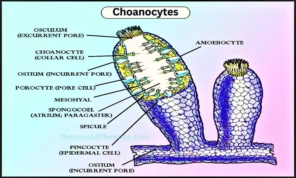
Choanocytes: Choanocytes are specialized cells found in sponges (Phylum Porifera) that play a crucial role in their feeding and filtration processes. Also known as collar cells, choanocytes are distinctive due to their unique morphology. Each choanocyte consists of a central cell body with a long, whip-like flagellum projecting outward. Flagella are enclosed by microvilli that give cells their unique appearance.
Choanocytes are typically located in the inner lining of a sponge’s body, forming a layer called the choanoderm. The collar cells are arranged in chambers or canals within the sponge, creating a complex network. The beating of the flagella generates water currents that bring in food particles and oxygen while removing waste and carbon dioxide.
The primary function of choanocytes is filter feeding. As water flows through the sponge, the collar cells trap suspended organic matter and planktonic organisms on their collars. These captured particles are then engulfed and digested by the choanocytes, contributing to the sponge’s nutrition. Choanocytes are also involved in sexual reproduction, producing sperm or eggs.
As well as their feeding role, choanocytes have attracted scientific interest due to their similarity to choanoflagellates – single-celled organisms considered close relatives of animals. Studying choanocytes provides insights into the evolutionary origins and transition from unicellular organisms to multicellular ones.
Pinacocytes: Pinacocytes are specialized cells found in sponges (Phylum Porifera) that form the outermost layer of the sponge’s body wall. Pinacocytes are flat, epithelial-like cells that cover the surface of the sponge and line its internal canals and chambers. These cells play essential roles in protection, support, and regulation within the sponge’s body.
The primary function of pinacocytes is to create a protective barrier for the sponge. They form a continuous layer that acts as a waterproof covering, preventing the entry of harmful substances and pathogens into the sponge’s body. Pinacocytes also help regulate the exchange of gases, nutrients, and waste products between the sponge and its environment.
They can control the flow of water through the sponge by contracting or relaxing, thus contributing to the overall water circulation and maintaining optimal conditions for the sponge’s survival.
In addition to their protective function, pinacocytes also provide structural support to the sponge. These cells are connected through intercellular junctions, forming a cohesive layer that reinforces the sponge’s body structure. By maintaining the integrity of the body wall, pinacocytes contribute to the overall stability and shape of the sponge.
Pinacocytes are involved in asexual reproduction in some sponge species. They undergo cell division and reproduce new cells which contribute to growth and regeneration within their bodies. Self-renewal and tissue regeneration play an integral part in ensuring survival and adaptation to changing environmental conditions for sponges.
Pinacocytes play crucial roles in the functioning of sponges, providing protection, and support, and maintaining the integrity of the sponge’s body. They work in conjunction with other cell types, such as choanocytes, to ensure the overall health and survival of the sponge organism.
Importance of choanocytes and pinacocytes in their respective organisms
Choanocytes and pinacocytes are both crucial cell types found in sponges (Phylum Porifera), and they serve essential functions within their respective organisms.
Their importance can be summarized as follows:
Choanocytes:
Feeding and Filtration: Choanocytes are primarily responsible for filter-feeding in sponges. Their collar structures trap and capture organic particles and plankton from the water, which are then engulfed and digested, providing the sponge with nutrients. Choanocytes play a vital role in the sponge’s ability to extract food from the surrounding environment.
Water Circulation: The beating of the flagella on choanocytes generates water currents within the sponge. These currents facilitate the flow of water through the sponge’s body, bringing in oxygen and food while removing waste and carbon dioxide. The coordinated action of choanocytes helps maintain a continuous flow of water, contributing to the sponge’s respiratory and metabolic processes.
Reproduction: Choanocytes are involved in sexual reproduction in sponges. Producing both sperm and eggs that can then be released into the water for fertilization purposes. Choanocytes are responsible for the production and release of gametes, contributing to the sponge’s reproductive cycle.
Pinacocytes:
Protection and Barrier Function: Pinacocytes form the outermost layer of the sponge’s body wall, serving as a protective barrier against harmful substances, pathogens, and physical damage. They create a waterproof covering that helps maintain the internal environment of the sponge and prevents the entry of unwanted materials.
Structural Support: Pinacocytes provide structural support to the sponge. By forming a cohesive layer and connecting through intercellular junctions, they contribute to the overall stability, shape, and integrity of the sponge’s body. Pinacocytes help maintain the structural framework of the sponge and ensure its proper functioning.
Regulation of Exchange: Pinacocytes play a role in regulating the exchange of gases, nutrients, and waste products between the sponge and its environment. They control the flow of water through the sponge by contracting or relaxing, thereby influencing the rate of gas exchange and the transport of nutrients and waste products.
Asexual Reproduction: In some sponge species, pinacocytes are involved in asexual reproduction. They can undergo cell division, contributing to the growth, repair, and regeneration of the sponge’s body. Pinacocytes possess the capacity for self-renewal, an ability that plays an essential role in an organism’s ability to adapt and recover from injuries or disturbances.
Choanocytes and pinacocytes are vital cell types that play distinct but complementary roles in the functioning of sponges. Choanocytes are specialized for filter-feeding, water circulation, and reproduction, while pinacocytes provide protection, support, and regulate exchange processes.
Together, these cell types contribute to the overall health, survival, and ecological success of sponges as essential components of their cellular organization and functionality.
What are Choanocytes?
Choanocytes, also known as collar cells, are specialized cells found in sponges (Phylum Porifera) and a few other organisms. They are named for their distinctive morphology, which includes a central cell body with a long, whip-like flagellum surrounded by a collar of microvilli.
Choanocytes play a crucial role in the feeding and filtration processes of sponges. They are primarily responsible for generating water currents and capturing food particles from the surrounding environment.
The beating of the flagellum creates a flow of water, which brings in nutrient-rich water and removes waste products. As water passes through the sponge, choanocytes use their collar structures to trap suspended organic matter and planktonic organisms. The captured particles are then engulfed and digested by the choanocytes, providing nutrients for the sponge.
The collar of microvilli surrounding the flagellum helps to increase the surface area available for trapping food particles. The microvilli create a sieve-like structure that allows the choanocytes to efficiently capture small organisms and organic particles from the water.
Apart from their feeding function, choanocytes also have significance in terms of evolution. They bear striking resemblances to choanoflagellates, considered one of the closest living relatives to animals. Choanocytes and choanoflagellates offer insight into the evolutionary roots and transition from unicellular organisms into multicellular ones.
In summary, choanocytes are specialized cells found in sponges that have a distinct structure involving a central cell body, a flagellum, and a collar of microvilli. They are primarily responsible for filter-feeding, generating water currents, and capturing food particles from the surrounding environment. Choanocyte research also contributes to our knowledge of multicellular organism evolution.
Structure of Choanocytes
The structure of choanocytes, also known as collar cells, is specialized for their unique functions in filter-feeding and generating water currents within sponges.
The main components of choanocytes include:
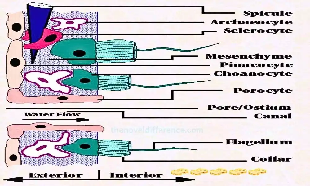
Cell Body: The choanocyte has a central cell body that contains the nucleus and other cellular organelles. The cell body is connected to the rest of the sponge’s body by a basal body or stalk.
Flagellum: Extending outward from the cell body is a long, whip-like structure called the flagellum. The flagellum is typically surrounded by a protective sheath called the collar.
Collar: The collar is a ring-like structure composed of microvilli, which are tiny, hair-like projections. The collar surrounds the base of the flagellum and extends outward. The microvilli create a sieve-like structure that increases the surface area available for capturing food particles.
Microvilli: The microvilli are specialized projections of the cell membrane that form the collar. They are lined with microfilaments and contain receptors that help in capturing food particles. The microvilli play a crucial role in filtering and trapping organic matter from the surrounding water.
The unique structure of choanocytes allows them to function effectively in filter-feeding. As water flows through the sponge, the beating of the flagella creates a flow that brings in water and suspended particles. The collar and microvilli trap and capture food particles from the water, which are then engulfed and digested by the choanocytes.
It is important to note that the exact structure of choanocytes can vary among different sponge species. The key features of a central cell body, flagellum, and collar with microvilli are generally present in most choanocytes.
What are Pinacocytes?
Pinacocytes are specialized cells found in sponges (Phylum Porifera) that make up the outermost layer of the sponge’s body wall. They are flat, epithelial-like cells that cover the surface of the sponge and line its internal canals and chambers. Pinacocytes play important roles in protection, support, and regulation within the sponge’s body.
The main characteristics and functions of pinacocytes are as follows:
Protective Barrier: Pinacocytes form a continuous layer that acts as a protective barrier for the sponge. They create a waterproof covering that helps prevent the entry of harmful substances, pathogens, and physical damage into the sponge’s body. The pinacoderm, formed by the layer of pinacocytes, serves as a barrier to maintain the internal environment of the sponge.
Structural Support: Pinacocytes contribute to the structural integrity of the sponge. They are tightly connected through intercellular junctions, forming a cohesive layer. This layer of pinacocytes reinforces the sponge’s body structure, providing support and maintaining the overall shape of the sponge. They help maintain the sponge’s physical integrity and stability.
Regulation of Exchange: Pinacocytes play a role in regulating the exchange of gases, nutrients, and waste products between the sponge and its environment. They control the flow of water through the sponge by contracting or relaxing, which influences the rate of gas exchange and the transport of nutrients and waste products. This regulation helps maintain the balance of essential substances within the sponge’s body.
Asexual Reproduction: In some sponge species, pinacocytes are involved in asexual reproduction. They can undergo cell division, contributing to the growth, repair, and regeneration of the sponge’s body. Pinacocytes can self-renew and differentiate into other cell types, enabling the sponge to generate new tissue and form new individuals through asexual reproduction.
Pinacocytes work in conjunction with other cell types, such as choanocytes and archaeocytes, to ensure the overall functioning of the sponge. Together, they form the cellular architecture of the sponge, playing vital roles in protection, support, regulation, and reproduction.
Structure of Pinacocytes
Pinacocytes are specialized cells found in sponges (Phylum Porifera) that make up the outermost layer of the sponge’s body wall. The structure of pinacocytes is adapted to their functions in protection, support, and regulation.
Here are the key structural features of pinacocytes:
Flat Epithelial Cells: Pinacocytes are flat, squamous cells that have a thin and flattened shape. Their morphology resembles that of epithelial cells found in other organisms. The flat structure allows pinacocytes to form a continuous layer and cover the surface of the sponge’s body, as well as line the internal canals and chambers.
Cell Membrane: The cell membrane forms the outer boundary of pinacocytes. It acts as a selective barrier, controlling the exchange of substances between the sponge’s internal environment and the surrounding water.
Intercellular Junctions: Pinacocytes are tightly connected through specialized intercellular junctions. These junctions help maintain the integrity of the pinacoderm, the layer formed by pinacocytes. The tight junctions between adjacent pinacocytes ensure the cohesion and stability of the cell layer, contributing to the structural integrity of the sponge.
Cytoplasm and Nucleus: The cytoplasm of pinacocytes contains various cellular organelles, including the nucleus. The nucleus contains the genetic material of the cell and plays a role in cellular activities such as gene expression and regulation.
Lack of Specialized Organelles: Unlike some other cell types, pinacocytes generally lack specialized organelles like flagella, cilia, or collar structures. Their primary function is not related to active movement or filtration but rather to protection, support, and regulation within the sponge’s body.
The structure of pinacocytes is characterized by their flat, epithelial-like shape, intercellular junctions, and lack of specialized organelles. These features allow pinacocytes to form a protective barrier, provide structural support, and participate in the regulation of exchange processes within the sponge’s body.
Difference Between Choanocytes and Pinacocytes
Choanocytes and pinacocytes are two distinct cell types found in sponges (Phylum Porifera) with different structures and functions.
Here are the key differences between choanocytes and pinacocytes:
Structure:
Choanocytes: Choanocytes have a distinctive structure that includes a central cell body with a long, whip-like flagellum surrounded by a collar of microvilli. The flagellum extends outward, while the collar surrounds its base.
Pinacocytes: Pinacocytes are flat, epithelial-like cells that cover the surface of the sponge’s body and line its internal canals and chambers. They have a thin and flattened shape, forming a continuous layer.
Location:
Choanocytes: Choanocytes are typically found in the inner lining of the sponge’s body, forming a layer called the choanoderm. They are often located within chambers or canals, creating a complex network.
Pinacocytes: Pinacocytes form the outermost layer of the sponge’s body wall. They cover the surface of the sponge and line its internal canals and chambers.
Function:
Choanocytes: Choanocytes are primarily involved in filter-feeding and generating water currents. They use their collar structures and flagella to trap and capture food particles from the surrounding water, while the beating of the flagella creates water currents for respiration and waste removal. Choanocytes also play a role in reproduction by producing gametes.
Pinacocytes: Pinacocytes have multiple functions related to protection, support, and regulation. They form a protective barrier against harmful substances and physical damage, provide structural support to the sponge’s body, regulate the exchange of gases, nutrients, and waste products, and may be involved in asexual reproduction.
Specialized Structures:
Choanocytes: Choanocytes have specialized structures, including the flagellum and collar with microvilli. These structures are adapted for filter-feeding and capturing food particles from the water.
Pinacocytes: Pinacocytes generally lack specialized structures like flagella or collars. Their structure is more focused on forming a continuous layer and providing protection and support to the sponge.
Role in Water Flow:
Choanocytes: Choanocytes actively contribute to water circulation by beating their flagella, generating water currents within the sponge. This helps bring in food particles and remove waste.
Pinacocytes: Pinacocytes do not actively contribute to water flow. They can regulate the flow of water by contracting or relaxing, influencing the rate of gas exchange and the transport of nutrients and waste products.
Choanocytes and pinacocytes differ in terms of their structure, location within the sponge, specialized structures, functions, and their role in water flow. Choanocytes are specialized for filter-feeding and water circulation within the sponge, while pinacocytes primarily provide protection, support, and regulate exchange processes.
Similarities between Choanocytes and Pinacocytes
While choanocytes and pinacocytes have distinct functions and structures.
There are a few similarities between these two cell types found in sponges:
- Both Found in Sponges: Choanocytes and pinacocytes are both cell types that are commonly found in sponges (Phylum Porifera). They are integral components of the sponge’s cellular organization.
- Importance in Sponge Functioning: Both choanocytes and pinacocytes play crucial roles in the overall functioning of sponges. While their specific functions differ, they contribute to the survival, growth, and overall health of the sponge organism.
- Embryonic Origin: Both choanocytes and pinacocytes are derived from the same embryonic cell layer, known as the pinacoderm. They differentiate into their specialized forms during the development of the sponge.
- Contribution to Sponge Physiology: Choanocytes and pinacocytes, along with other cell types, work together to maintain the physiological processes within the sponge. They contribute to feeding, gas exchange, waste removal, and reproduction, which are essential for the sponge’s survival.
- Multicellularity: Both choanocytes and pinacocytes are part of the multicellular structure of sponges. They demonstrate the cellular specialization and cooperation necessary for the functioning of complex multicellular organisms.
It is important to note that while there are some similarities between choanocytes and pinacocytes, they have distinct structures, locations, and functions within the sponge. These similarities highlight the diverse and specialized cellular components that contribute to the overall organization and functioning of sponges.
Examples from Organisms
Examples of organisms that possess choanocytes and pinacocytes are primarily found within the Phylum Porifera, which consists of sponges.
Here are examples of organisms where choanocytes and pinacocytes can be found:
Choanocytes:
- Sponges (Phylum Porifera): Choanocytes are the defining characteristic of sponges. They are present in all classes of sponges, including Calcarea, Hexactinellida, and Demospongiae. Examples of sponges that possess choanocytes include the common bath sponge (Spongilla), vase sponge (Callyspongia), and barrel sponge (Xestospongia).
- Choanoflagellates: While not part of the animal kingdom, choanoflagellates are single-celled organisms that are the closest living relatives of animals. They have choanocyte-like cells that resemble the choanocytes found in sponges. Choanoflagellates are found in various aquatic environments.
Pinacocytes:
- Sponges (Phylum Porifera): Pinacocytes are also present in all classes of sponges. They make up the outermost layer of the sponge’s body wall and are found in species such as the common bath sponge (Spongilla), sea sponge (Porifera), and glass sponge (Hexactinellida).
It’s worth noting that while choanocytes are predominantly found in sponges and choanoflagellates, pinacocytes are exclusive to sponges within the animal kingdom. These examples illustrate the presence of choanocytes and pinacocytes in various sponge species, highlighting their significance in the functioning of these organisms.
Conclusion
Choanocytes and pinacocytes are two distinct cell types found in sponges (Phylum Porifera) that contribute to the overall functioning and survival of these organisms. Choanocytes are specialized cells responsible for filter-feeding, generating water currents, and reproduction.
They have a unique structure with a central cell body, a flagellum, and a collar of microvilli. Choanocytes are found in the inner lining of the sponge’s body and play a crucial role in capturing food particles and maintaining water circulation.