 by
by Two commonly used techniques for studying chromosomes are chromosome banding and chromosome painting. While both methods involve the visualization of chromosomes, they differ in their approach and the information they provide. We will explore the difference between chromosome banding and chromosome painting, highlighting their unique features, applications, and significance in genetic research.
Definition of Chromosome Banding and Chromosome Painting
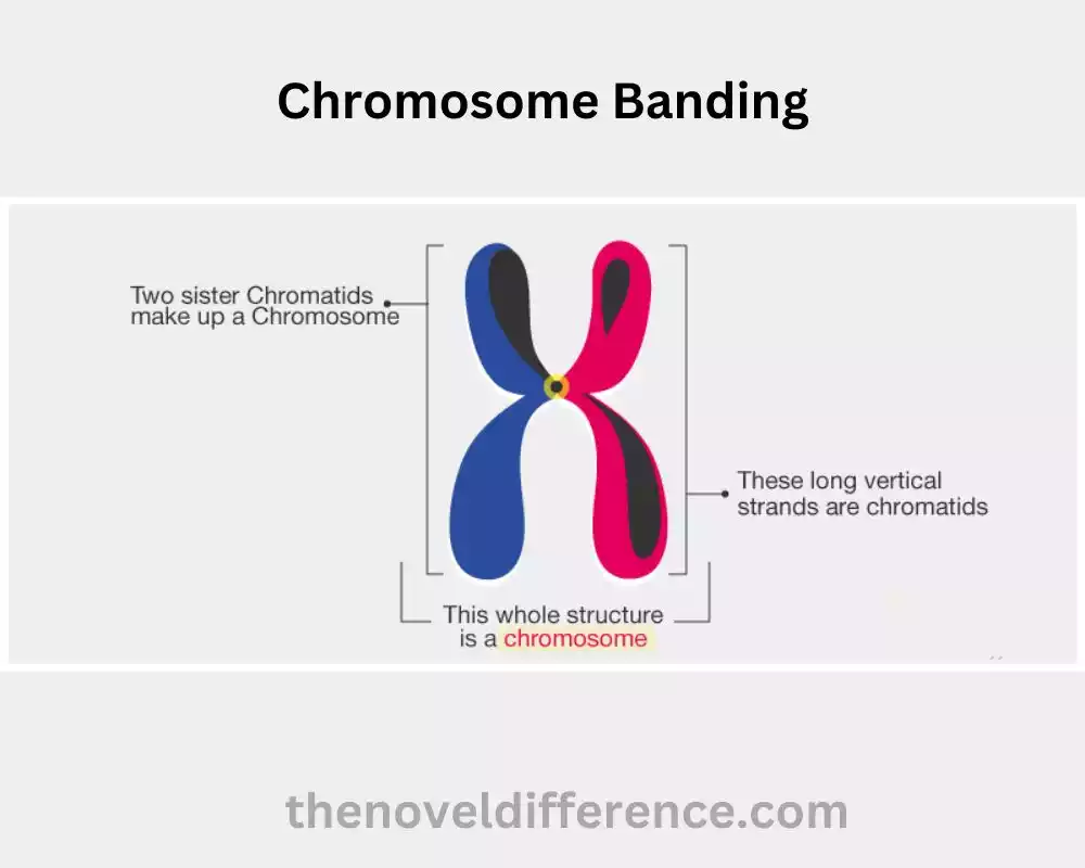
Chromosome Banding: Chromosome banding alludes to a procedure utilized in cytogenetics to recolor and distinguish particular districts of chromosomes. It involves the application of specific dyes or chemical treatments to chromosomes, which results in distinct banding patterns along the length of the chromosomes. These banding patterns are unique to each chromosome and provide a way to visualize and differentiate between individual chromosomes.
Different banding strategies, such as G-banding, Q-banding, R-banding, and C-banding, are utilized to form characteristic banding designs on chromosomes. Chromosome banding is broadly utilized for different applications, counting the distinguishing proof of chromosomal variations from the norm, mapping particular qualities or hereditary markers, and considering developmental connections between species.
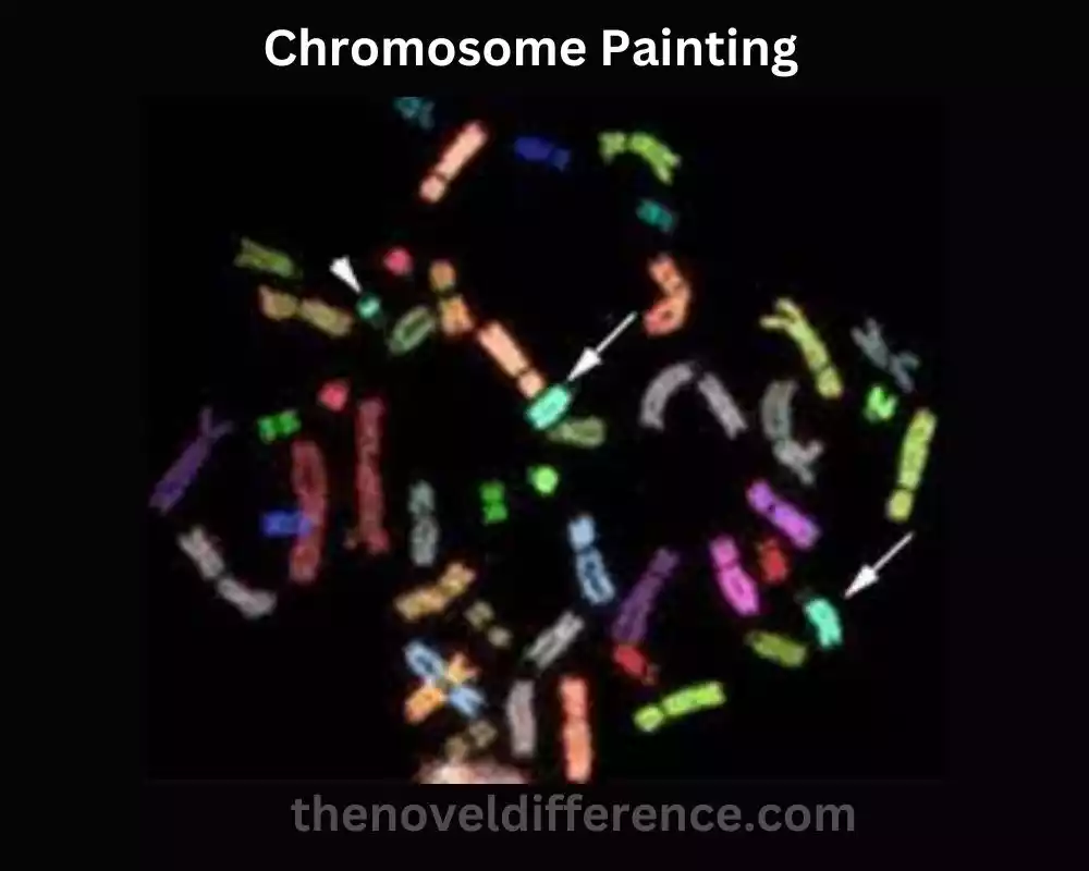
Chromosome Painting: Chromosome portrayal, moreover known as chromosome-specific or whole-chromosome portrayal, could be a cytogenetic method utilized to imagine and distinguish particular chromosomes or chromosomal districts. It involves the use of fluorescently labeled probes that hybridize into complementary sequences on the chromosomes of interest. These probes are typically derived from whole chromosomes or specific regions of chromosomes and are labeled with different fluorochromes to produce distinct colors for each chromosome or chromosomal segment.
By using fluorescence in situ hybridization (FISH) or other related techniques, chromosome painting allows researchers to visualize the spatial arrangement, structural abnormalities, and interactions of chromosomes within the cell nucleus. It is especially valuable within the location of chromosomal improvements, translocations, and the consideration of chromosomal advancement and phylogenetics. Chromosome painting has important applications in clinical genetics, research, and reproductive medicine.
Importance of studying chromosome structure and function
Considering chromosome structure and work is of foremost significance in different areas of science and pharmaceutical.
Here are some key reasons why the investigation of chromosome structure and function is significant:
1. Understanding Genetic Material: Chromosomes are the carriers of genetic material in living organisms. They comprise DNA, which contains the information essential for the advancement, development, and working of all life forms. By studying chromosome structure and function, scientists can gain insights into the organization, packaging, and expression of genes, providing a foundation for understanding the fundamental principles of genetics.
2. Identification of Genetic Disorders: Many genetic disorders and diseases are caused by abnormalities or mutations in chromosomes. Analyzing chromosome structure permits the discovery of chromosomal distortions, such as erasures, duplications, reversals, and translocations, which can lead to hereditary disarranges. By studying these abnormalities, researchers can identify the genetic basis of diseases and develop diagnostic tests for early detection and intervention.
3. Prenatal Diagnosis and Genetic Counseling: Chromosome analysis plays a crucial role in prenatal diagnosis, enabling the identification of chromosomal abnormalities in developing fetuses. Procedures like amniocentesis and chorionic villus testing (CVS) permit the collection of fetal cells for chromosomal investigation, supporting within the early location of conditions like Down disorder, Turner disorder, and other chromosomal disarranges. This information enables genetic counseling, which helps parents make informed decisions regarding the management and care of their unborn child.
4. Evolutionary Studies: Comparative cytogenetics, which involves comparing chromosome structure and organization across different species, provides insights into evolutionary relationships and genetic diversity. By analyzing chromosome structure, analysts can think about chromosomal modifications, changes in chromosome number, and other developmental occasions, contributing to our understanding of speciation, versatile radiation, and the advancement of genomes.
5. Cancer Research: Chromosome abnormalities are commonly associated with cancer. Considering the chromosomal changes in cancer cells can offer assistance recognize oncogenes (qualities related to cancer improvement) and tumor silencer qualities (qualities that hinder tumor arrangement). Chromosome examination can help in cancer determination, guess, and the advancement of focused treatments.
6. Assisted Reproductive Technologies: Chromosome analysis is vital in assisted reproductive technologies, such as in vitro fertilization (IVF). Preimplantation hereditary testing includes the examination of embryos for chromosomal variations from the norm sometime recently they are embedded into the uterus, expanding the chances of effective pregnancies and lessening the hazard of hereditary disarranges.
Considering chromosome structure and work is fundamental for understanding the hereditary premise of infections, diagnosing hereditary disarranges, unraveling developmental connections, progressing cancer inquiries, and progressing regenerative innovations. It provides valuable insights into the organization and functioning of genetic material, leading to advancements in various fields of biology and medicine.
What is Chromosome Banding?
Chromosome banding is a cytogenetic technique used to stain and visualize specific regions of chromosomes. It involves the treatment of chromosomes with dyes or chemicals that create distinct banding patterns along the length of the chromosomes. These banding patterns help in the identification and differentiation of individual chromosomes and specific chromosomal regions.
Different banding techniques have been developed, each producing characteristic banding patterns.
The most commonly used banding techniques include:
1. G-banding (Giemsa banding): This technique involves staining chromosomes with the dye Giemsa, which produces a pattern of light and dark bands along the length of the chromosomes. G-banding is broadly utilized in karyotyping and permits the recognizable proof of person chromosomes and the discovery of chromosomal anomalies.
2. Q-banding (Quinacrine banding): Quinacrine mustard is a fluorescent dye that selectively binds to regions of DNA rich in adenine and thymine. Q-banding produces a pattern of fluorescent bands on chromosomes, which can be visualized using fluorescence microscopy. Q-banding is particularly useful in identifying certain chromosomal abnormalities, such as fragile sites.
3. R-banding (Reverse banding): R-banding is the reverse pattern of G-banding, where the dark bands appear light, and the light bands appear dark. This technique involves pretreating chromosomes with heat or proteolytic enzymes before staining. R-banding is valuable for studying chromosomal breakpoints and rearrangements.
4. C-banding (Constitutive heterochromatin banding): C-banding is used to stain the constitutive heterochromatin regions of chromosomes. Heterochromatin is a densely packed, transcriptionally inactive DNA region, often found near the centromeres and telomeres of chromosomes. C-banding reveals the distribution and structure of constitutive heterochromatin, aiding in the identification of specific chromosome regions.
Chromosome banding strategies give analysts a visual representation of chromosome structure, permitting the recognizable proof of person chromosomes, location of chromosomal anomalies, mapping of particular qualities or hereditary markers, and comparative genomic ponders. By looking at the banding designs, researchers can pick up experiences in the organization, course of action, and working of chromosomes, contributing to our understanding of hereditary qualities, advancement, and human well-being.
Techniques used in chromosome banding
Several techniques are used in chromosome banding to stain and differentiate specific regions of chromosomes.
The commonly employed techniques include:
1. G-banding (Giemsa banding): G-banding is one of the most widely used chromosome banding techniques. It involves staining chromosomes with the dye Giemsa after treating them with a mild proteolytic enzyme, such as trypsin. The chromosomes are then visualized under a microscope. G-banding produces a characteristic pattern of alternating light and dark bands along the length of the chromosomes, allowing for the identification and differentiation of individual chromosomes.
2. Q-banding (Quinacrine banding): Q-banding utilizes the fluorescent dye quinacrine mustard, which selectively binds to regions of DNA rich in adenine and thymine (AT-rich regions). Chromosomes are stained with quinacrine mustard and examined under fluorescence microscopy. This technique produces a distinct pattern of fluorescent bands on the chromosomes, with AT-rich regions appearing bright. Q-banding is particularly useful for identifying fragile sites and certain chromosomal abnormalities.
3. R-banding (Reverse banding): R-banding is the reverse pattern of G-banding, where the dark bands appear light, and the light bands appear dark. This technique involves pretreating chromosomes with heat or proteolytic enzymes before staining with Giemsa. R-banding can reveal specific chromosomal breakpoints and rearrangements, providing additional information compared to G-banding.
4. C-banding (Constitutive heterochromatin banding): C-banding is used to stain the constitutive heterochromatin regions of chromosomes. Constitutive heterochromatin consists of repetitive DNA sequences and is typically found near the centromeres and telomeres of chromosomes. C-banding includes treating chromosomes with soluble bases or other denaturing operators, taken after by recoloring with a DNA-specific color, such as propidium iodide or DAPI (4′,6-diamidino-2-phenylindole). Constitutive heterochromatin regions appear intensely stained, while euchromatic regions remain relatively unstained. C-banding helps identify specific chromosomal regions and structural variations.
These procedures, collectively known as chromosome banding, give unmistakable designs and varieties within the recoloring of chromosomes, permitting the recognizable proof, characterization, and investigation of person chromosomes and particular chromosomal locales. They are important apparatuses in cytogenetic inquiry about, clinical hereditary qualities, and demonstrative applications, empowering the location of chromosomal variations from the norm, hereditary disarranges, and chromosomal modifications.
Giemsa banding (G-banding)
Giemsa banding, also known as G-banding, is a widely used chromosome banding technique that helps visualize and differentiate individual chromosomes. G-banding involves staining chromosomes with the dye Giemsa, which selectively binds to the DNA and reveals characteristic banding patterns.
Here’s a brief diagram of the G-banding strategy:
1. Chromosome Preparation: The primary step in G-banding is the planning of chromosomes. This typically involves obtaining cells from a tissue sample, culturing them in the laboratory, and arresting them at a specific stage of the cell cycle (often metaphase) when the chromosomes are condensed and visible.
2. Fixation: The cells are at that point treated with a fixative, such as methanol and acidic corrosive, to protect the chromosome structure.
3. Proteolytic Treatment: To prepare the chromosomes for staining, a proteolytic enzyme, such as trypsin, is applied to remove some of the proteins associated with the chromosomes, allowing better penetration of the dye.
4. Staining with Giemsa: The chromosomes are stained with Giemsa dye, which consists of a mixture of azure and eosin. Giemsa dye binds to the DNA, resulting in differential staining along the chromosomes.
5. Banding Pattern Visualization: The stained chromosomes are examined under a microscope equipped with appropriate magnification and lighting conditions. The Giemsa-stained chromosomes display a pattern of alternating light (G-negative) and dark (G-positive) bands along their length. The bands correspond to specific regions of the chromosomes and are numbered from the centromere outward.
6. Karyotyping: The banding patterns are analyzed, and the chromosomes are matched to create a karyotype, which is a visual representation of the complete set of chromosomes of an individual or a species. The karyotype allows for the identification and arrangement of chromosomes according to their size, centromere position, and banding patterns.
G-banding provides a reproducible and standardized way to analyze chromosome structure and identify individual chromosomes. The banding patterns revealed by Giemsa staining allow for the detection of chromosomal abnormalities, such as deletions, duplications, translocations, and inversions. G-banding is widely used in clinical cytogenetics for the diagnosis of genetic disorders and in research settings for studying chromosomal abnormalities, evolutionary relationships, and genomic organization.
Quinacrine banding (Q-banding)
Quinacrine banding, also known as Q-banding, is a chromosome banding technique that uses the fluorescent dye quinacrine mustard to stain and visualize specific regions of chromosomes. Q-banding is particularly useful in identifying certain chromosomal abnormalities and fragile sites.
Here’s a brief overview of the Q-banding technique:
1. Chromosome Preparation: Similar to other chromosome banding techniques, the first step in Q-banding involves obtaining cells from a tissue sample, culturing them, and arresting them at a suitable stage of the cell cycle when the chromosomes are condensed and visible.
2. Fixation: The cells are treated with a fixative to preserve the chromosome structure.
3. Quinacrine Staining: Quinacrine mustard, a fluorescent dye, is applied to the chromosomes. Quinacrine selectively binds to adenine-thymine (AT)-rich regions of DNA. These AT-rich regions are often found in constitutive heterochromatin regions near the centromeres and the short arms of acrocentric chromosomes.
4. Visualization: The stained chromosomes are examined using fluorescence microscopy. Quinacrine-stained regions appear bright and emit a yellow-green fluorescence under ultraviolet (UV) light.
5. Banding Pattern Interpretation: The Q-banding pattern observed under the microscope reveals the distribution of AT-rich regions along the chromosomes. The specific regions that appear bright correspond to the AT-rich regions of the chromosomes. The banding pattern can help identify certain chromosomal abnormalities, such as fragile sites or regions prone to breakage.
Q-banding is useful in identifying fragile sites, which are specific chromosomal regions that are susceptible to breakage under certain conditions. These fragile sites may be associated with genetic disorders or predispose individuals to chromosomal rearrangements. Q-banding can also aid in identifying certain chromosomal abnormalities and understanding the structural organization of chromosomes.
It’s critical to note that Q-banding is less commonly utilized compared to other banding methods, such as G-banding. It can be a valuable addition to the cytogenetic toolkit, especially in specific research or diagnostic situations where the identification of AT-rich regions or fragile sites is of interest.
What is Chromosome Painting?
Chromosome portrayal, too known as chromosome-specific or whole-chromosome portrayal, could be a cytogenetic method utilized to imagine and recognize particular chromosomes or chromosomal locales. It involves the use of fluorescently labeled probes that hybridize into complementary sequences on the chromosomes of interest.
Here’s an overview of the chromosome painting technique:
1. Probe Preparation: To perform chromosome painting, specific DNA probes are prepared. These probes are derived from whole chromosomes or specific regions of chromosomes. The tests are labeled with fluorescent colors that emanate diverse colors, such as green, ruddy, or blue. Each chromosome or chromosomal region is typically labeled with a unique color.
2. Chromosome Preparation: Cells containing the chromosomes of interest are collected and prepared for analysis. This may involve culturing cells, arresting them at a specific stage of the cell cycle, and fixing them to preserve the chromosome structure.
3. Probe Hybridization: The labeled DNA probes are applied to the prepared chromosomes and allowed to hybridize (bind) to their complementary DNA sequences on the chromosomes. The hybridization process typically occurs under controlled temperature and humidity conditions to ensure optimal probe binding.
4. Visualization: The hybridized chromosomes are examined under a fluorescence microscope equipped with appropriate filters to detect the fluorescent signals emitted by the labeled probes. Each chromosome or chromosomal region appears as a distinct fluorescent color, allowing for the visualization and identification of individual chromosomes or specific chromosomal regions.
5. Analysis and Interpretation: The resulting images are analyzed to determine the presence, location, and patterns of the fluorescent signals on the chromosomes. This data can be utilized to recognize particular chromosomes, distinguish chromosomal modifications, explore auxiliary anomalies, and think about chromosomal advancement or phylogenetics.
Chromosome painting offers several advantages in cytogenetic analysis. It allows for the visualization of chromosomes in their entirety or specific regions of interest, overcoming the limitations of traditional banding techniques. It provides a comprehensive view of chromosomal organization and interactions within the cell nucleus. Chromosome painting is particularly valuable in the detection of chromosomal rearrangements, translocations, and the identification of marker chromosomes. It is widely used in research, clinical genetics, and reproductive medicine for applications such as cancer cytogenetics, prenatal diagnosis, and the study of evolutionary relationships among species.
Chromosome portrayal could be a capable apparatus that helps with the visualization, distinguishing proof, and investigation of chromosomes, contributing to our understanding of the chromosomal structure, work, and hereditary differences.
Procedure and Principles of Chromosome Painting
The procedure of chromosome painting involves several steps and follows specific principles.
Here’s a general overview of the procedure and principles of chromosome painting:
Procedure:
1. Chromosome Preparation: Cells containing the chromosomes of interest are collected and prepared for analysis. This typically involves culturing cells, arresting them at a specific stage of the cell cycle, and fixing them to preserve the chromosome structure. Different cell types and culture conditions may be used depending on the specific study objectives.
2. Probe Preparation: Specific DNA probes are prepared for the chromosomes or chromosomal regions of interest. These probes are typically derived from whole chromosomes or specific regions of chromosomes. The tests are labeled with fluorescent colors, such as green, ruddy, or blue, which transmit unmistakable colors when visualized beneath a fluorescence magnifying lens.
3. Probe Hybridization: The labeled DNA probes are applied to the prepared chromosomes and allowed to hybridize (bind) to their complementary DNA sequences on the chromosomes. The hybridization process is facilitated by incubating the chromosomes and probes together under controlled temperature and humidity conditions. This allows for specific binding between the probes and their target sequences on the chromosomes.
4. Wash and Counterstain: After the hybridization step, the excess, unbound probes are washed away to reduce background noise and improve the specificity of the signal. A counterstain, such as DAPI (4′,6-diamidino-2-phenylindole), may be applied to visualize the overall chromosomal morphology and aid in locating specific chromosomes or regions.
5. Visualization and Analysis: The hybridized chromosomes are examined under a fluorescence microscope equipped with appropriate filters to detect the fluorescent signals emitted by the labeled probes. Each chromosome or chromosomal region appears as a distinct fluorescent color, allowing for the visualization and identification of individual chromosomes or specific chromosomal regions. The resulting images can be captured and analyzed using specialized software for further interpretation and analysis.
Principles:
1. Specific Hybridization: The labeled DNA probes used in chromosome painting are designed to bind specifically to their complementary DNA sequences on the chromosomes. This ensures that the probes hybridize only to the target chromosomes or regions of interest, providing accurate identification and visualization.
2. Unique Colors: Each chromosome or chromosomal region is typically labeled with a unique fluorescent color. This principle enables the differentiation and identification of individual chromosomes or regions within the chromosome complement.
3. Complementary DNA Binding: The labeled probes bind to their complementary DNA sequences on the chromosomes through base pairing. The degree of hybridization reflects the similarity and complementarity between the probes and their target sequences.
4. Visualization and Analysis: The fluorescence signals emitted by the labeled probes are detected and visualized using a fluorescence microscope. The resulting images are analyzed to determine the presence, location, and patterns of the fluorescent signals on the chromosomes. This analysis can aid in identifying specific chromosomes, detecting chromosomal rearrangements, and studying chromosomal organization and evolution.
By following these procedures and principles, chromosome painting allows for the visualization, identification, and analysis of chromosomes or specific chromosomal regions. It gives important bits of knowledge into the chromosomal structure, work, and hereditary variety, contributing to different regions of inquiry about and clinical applications.
Comparison Chart
Here’s a comparison chart highlighting the key differences between chromosome banding and chromosome painting:
| Chromosome Banding | Chromosome Painting |
|---|---|
| Staining chromosomes with dyes or chemicals to produce characteristic banding patterns along chromosomes | Using fluorescently labeled DNA probes that hybridize to specific chromosomes or chromosomal regions |
| Patterns of alternating light and dark bands along chromosomes | Fluorescent colors emitted by labeled probes on chromosomes |
| High-resolution visualization of chromosomal bands | Lower resolution compared to banding techniques |
| Diagnosis of chromosomal disorders, structural abnormalities, and research on chromosomal evolution and genome organization | Identification of chromosomal rearrangements, translocations, specific abnormalities, cancer cytogenetics, prenatal diagnosis, and comparative genomic studies |
| Precise mapping of chromosomal abnormalities and rearrangements | The broader view of chromosomal organization and interactions |
| Individual chromosomes and specific regions | Specific chromosomes or regions |
| G-banding, R-banding, C-banding, Q-banding | Chromosome-specific or whole-chromosome painting |
Conclusion
Chromosome banding and chromosome painting are valuable techniques in genetic research, offering unique insights into chromosomal structure, gene mapping, and the identification of chromosomal abnormalities. While chromosome banding provides an overview of the chromosomal organization, chromosome painting allows for the visualization of specific DNA sequences. By leveraging the strengths of these techniques, researchers can unravel the complexities of the genome, advancing our understanding of genetic disorders, cancer biology, and evolutionary relationships.