 by
by When it comes to studying cells, two important terms that often come up are total cell count and viable cell count. These terms play a significant part in different logical and therapeutic areas, counting microbiology, biotechnology, and cell culture. Understanding the difference between total cell count and viable cell count is essential for accurate data interpretation and experimental results. We’ll dive into the points of interest of these two concepts, clarifying their definitions, noteworthiness, and how they vary from each other.
Definition of Total Cell Count and Viable Cell Count
Total Cell Count: Total cell count alludes to the estimation of all cells displayed in a given test, notwithstanding their reasonability or utilitarian status. It gives data around the overall number of cells, counting both live and dead cells, as well as any cellular flotsam and jetsam or contaminants which will be displayed. The add-up-to-cell number is regularly communicated as the number of cells per unit volume (e.g., cells/mL) or as an add-up to the cell number for the whole test.
Viable Cell Count: Viable cell count specifically focuses on determining the number of live, viable cells within a sample. It avoids non-viable cells, such as dead cells or cells that have misplaced their capacity to isolate and work legitimately. Reasonable cell check is critical since it gives experiences into the well-being and usefulness of cells, which is significant in different applications such as cell culture, medicate revelation and cell-based treatments. Reasonable cell check is ordinarily communicated as the number of reasonable cells per unit volume (e.g., practical cells/mL).
Importance of cell counting in various fields
Cell counting plays a crucial role in various fields and disciplines due to its significance in understanding cellular behavior, assessing cell health, and facilitating research and applications.
The importance of cell counting can be observed in the following fields:
1. Biomedical Research: Cell counting is fundamental in biomedical research for studying cellular processes, identifying cell populations, and investigating disease mechanisms. It enables researchers to quantify changes in cell numbers, monitor cell proliferation or apoptosis, and evaluate the effects of experimental treatments or interventions.
2. Clinical Diagnostics: Cell counting is essential in clinical diagnostics for diagnosing diseases, monitoring patient conditions, and evaluating treatment responses. It makes a difference in deciding blood cell tallies (ruddy blood cells, white blood cells, platelets), evaluating resistant framework work, recognizing irregular cell populaces (e.g., cancer cells), and checking irresistible illnesses.
3. Bioprocessing and Cell Culture: Accurate cell counting is crucial for maintaining optimal cell growth and productivity. It aids in monitoring cell viability, assessing cell density for scaling up or down culture volumes, optimizing growth conditions, and determining the appropriate seeding density for experimental or production purposes.
4. Drug Discovery and Development: Cell counting is integral to drug discovery and development processes. It assists in assessing the cytotoxicity or efficacy of drug candidates, evaluating cell-based assays for screening potential drugs, optimizing drug dosages, and studying drug mechanisms of action.
5. Cell-Based Therapies and Tissue Engineering: Cell counting is vital in cell-based therapies and tissue engineering to ensure the quality, potency, and safety of cellular products. It helps in determining cell concentrations for transplantation, evaluating cell viability post-processing, monitoring cell expansion or differentiation, and assessing the impact of culture conditions on cellular behavior.
6. Microbiology and Microbial Ecology: Cell counting is critical in microbiology and microbial ecology studies. It aids in enumerating microbial populations, assessing bacterial or fungal growth, monitoring microbial contamination in food or water samples, and studying microbial community dynamics in various environments.
7. Environmental Monitoring: Cell counting is employed in environmental monitoring to evaluate the health and abundance of organisms in ecosystems. It helps in evaluating phytoplankton or zooplankton populations, evaluating microbial differences in soil or water tests, and checking the effect of poisons or natural changes on natural communities.
Cell counting plays a pivotal role in numerous fields, providing valuable insights into cellular dynamics, health, and interactions. It enables researchers and professionals to make informed decisions, advance scientific understanding, and develop applications and therapies for various purposes.
What is the Total Cell Count?
Total cell count alludes to the evaluation of all cells display in a given test, in any case of their reasonability or utilitarian status. It includes counting all cells, counting both live and dead cells, as well as any cellular flotsam and jetsam or contaminants which will be displayed. Total cell count provides information about the overall number of cells within a sample, offering a broad understanding of cell density or concentration.
There are various methods available to determine the total cell count, including manual techniques and automated systems. One common manual method involves using a hemocytometer or counting chamber, where cells are visually counted under a microscope within a defined area. Automated cell counters, on the other hand, utilize image analysis or flow cytometry to provide rapid and accurate total cell counts.
Total cell count is employed in various applications and fields, such as cell culture, biomedical research, clinical diagnostics, and environmental monitoring. It serves as a foundational parameter for assessing cell growth, evaluating treatment responses, quantifying cellular populations, and providing baseline information for further analyses.
Flow cytometry
Flow cytometry is a powerful analytical technique used to measure and analyze the characteristics of individual cells or particles in a heterogeneous sample. It empowers the synchronous examination of numerous parameters, such as cell estimate, shape, granularity, and the expression of particular particles on the cell surface or inside the cell.
The flow cytometry process involves the following steps:
1. Sample Preparation: The sample containing cells or particles of interest is prepared by appropriately treating, labeling, and suspending them in a fluid medium. Various labeling techniques can be employed, including fluorescent dyes, antibodies, or fluorescent proteins, to target specific cellular components or markers.
2. Sample Introduction: The prepared sample is introduced into the flow cytometer instrument, which consists of a fluidic system and a laser-based detection system. The sample is injected into a stream of sheath fluid, which creates a narrow, single-file flow of cells through a flow cell or cuvette.
3. Single-Cell Analysis: As the cells flow in a single-file manner, they pass through a laser beam(s) within the flow cell. The laser(s) excites the fluorochrome-labeled cells, causing them to emit fluorescence at specific wavelengths. Detectors capture the emitted fluorescence and convert it into electrical signals.
4. Signal Detection: The emitted fluorescence signals are detected by photomultiplier tubes (PMTs) or avalanche photodiodes (APDs) and are processed by electronic circuitry. Each detector corresponds to a specific fluorescence channel, allowing simultaneous measurement of multiple fluorescence parameters.
5. Data Acquisition and Analysis: The electronic signals representing the fluorescence parameters of individual cells are collected and recorded by a computer system. Advanced software is used to analyze the data, gating on specific cell populations, determining cell characteristics, and calculating various parameters such as cell count, cell viability, protein expression levels, and cell cycle distribution.
Flow cytometry offers several advantages, including high-speed analysis, multiparametric measurements, and the ability to analyze large cell populations quickly. It is broadly utilized in different areas, counting immunology, hematology, cancer investigate, stem cell science, microbiology, and sedate revelation. Applications of stream cytometry incorporate immunophenotyping, cell cycle examination, apoptosis discovery, intracellular protein examination, DNA substance investigation, and utilitarian measures, among others.
Automated cell counters
Automated cell counters are laboratory instruments used for the rapid and accurate quantification of cells in a sample. These devices automate the cell counting process, eliminating the need for manual counting using a hemocytometer or other traditional methods. Automated cell counters are commonly employed in research laboratories, clinical settings, bioprocessing facilities, and other areas where efficient and precise cell counting is essential.
The main features and functionalities of automated cell counters include:
1. Sample Handling: Automated cell counters typically require a small volume of the sample, which is loaded into the instrument through a dedicated sample chamber or cartridge. The test may be a suspension of cells or a real liquid, such as blood or cerebrospinal liquid.
2. Cell Detection and Counting: Once the sample is loaded, the automated cell counter employs various techniques to detect and count cells. These methods may incorporate optical strategies (e.g., light scrambling, retention, or fluorescence) or electrical impedance-based strategies. The instrument measures individual cells passing through a detection zone and records their counts.
3. Data Acquisition and Analysis: Automated cell counters capture and process the data obtained during the counting process. They employ built-in algorithms and software to differentiate cells from debris or artifacts, calculate total cell counts, and provide statistical analyses of the results.
4. Parameters and Assays: Many automated cell counters offer additional parameters and assays. These can include cell viability assessment (e.g., distinguishing live and dead cells), cell sizing, cell morphology analysis, concentration determination, and even cell sorting capabilities in more advanced systems.
5. User Interface: Automated cell counters have user-friendly interfaces that allow users to input settings, monitor the counting process, and access the generated results. They often include touchscreens, software interfaces, and data export options.
The preferences for utilizing robotized cell counters incorporate speed, precision, reproducibility, and the capacity to handle expansive numbers of tests proficiently. They reduce human error and subjectivity associated with manual counting methods, and they provide rapid results, which is especially beneficial in high-throughput applications.
Automated cell counters discover applications in a wide run of areas, such as cell culture, clinical diagnostics, investigative research facilities, pharmaceutical advancement, and bioprocessing. They are used for cell-based assays, monitoring cell growth, determining cell concentrations, evaluating treatment responses, and conducting quality control in various cellular analysis workflows.
What is a Viable Cell Count?
Viable cell count refers to the quantification of live, viable cells within a sample while excluding non-viable or dead cells. It particularly centers on deciding the number of cells that can preserve their physiological capacities, multiply, and contribute to natural forms.
Viable cell count is crucial in various applications where the viability and functionality of cells are important considerations, such as cell culture, cell-based therapies, tissue engineering, and drug development. It provides valuable information about the health, viability, and quality of the cell population under investigation.
Methods commonly used for viable cell counting include:
1. Colony-Forming Unit (CFU) Assay: This method involves diluting the cell suspension and plating it onto a culture medium that supports cell growth. After an incubation period, viable cells form colonies, which can be counted and used to determine the initial viable cell count.
2. Trypan Blue Exclusion Assay: Trypan blue is a dye that selectively stains non-viable cells with compromised cell membranes. In this assay, cells are mixed with trypan blue, and viable cells with intact membranes exclude the dye. Viable and non-viable cells are then counted using a hemocytometer or an automated cell counter, and the viable cell count is determined.
3. Fluorescent Dyes: Various fluorescent dyes, such as propidium iodide (PI), are used to distinguish between viable and non-viable cells. These dyes can penetrate cells with compromised membranes (non-viable cells) and emit fluorescence upon binding to nucleic acids. Flow cytometry or fluorescence microscopy is commonly employed to perform viable cell counting using fluorescent dyes.
4. Flow Cytometry-based Viability Assays: Flow cytometry allows for the assessment of cell viability using combinations of fluorescent dyes and specific markers. For instance, dual staining with fluorescent dyes such as propidium iodide (PI) and a fluorescently labeled viability marker (e.g., calcein-AM) enables the discrimination of live, dead, and apoptotic cells.
Accurate viable cell counting is essential in maintaining cell cultures, evaluating cell therapies’ potency and safety, assessing the impact of experimental conditions on cell viability, and determining the appropriate cell concentration for downstream applications. It provides critical information for optimizing cell growth, evaluating cell health, and ensuring the success of various cellular-based processes and therapies.
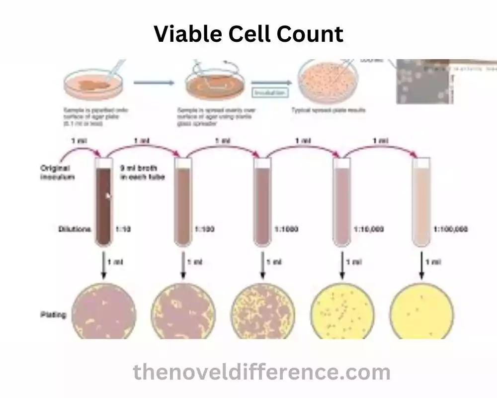
Colony-forming unit (CFU) assay
The Colony-Forming Unit (CFU) assay is a widely used method for determining the viable cell count in a sample. It is especially valuable when managing cells that can multiply and frame colonies, such as certain sorts of stem cells, microscopic organisms, and fungi.
The CFU assay involves the following steps:
1. Sample Dilution: The sample containing cells is serially diluted in a sterile medium to obtain dilutions that will allow for the formation of distinct colonies. This step helps ensure that the colonies are countable and not overcrowded.
2. Plating: Each dilution is plated onto a solid culture medium, such as agar, that provides a suitable environment for cell growth. The medium may contain nutrients, growth factors, or specific supplements to support the growth of the cells of interest.
3. Incubation: The agar plates are then incubated under appropriate conditions, such as temperature, humidity, and gas composition, to promote cell proliferation. The brooding period changes depending on the cell sort and wanted colony estimate but as a rule, ranges from a few hours to a few weeks.
4. Colony Formation: Viable cells in the sample divide and proliferate to form visible colonies on the agar surface. Each colony originates from a single viable cell or a cluster of cells, and each colony represents a colony-forming unit (CFU).
5. Colony Counting: After the incubation period, the plates are examined, and the colonies are counted. Careful attention is given to distinguishing individual colonies and excluding overlapping or merged colonies. Depending on the estimate and morphology of the colonies, manual checking can be performed employing a stereomicroscope, or robotized colony counters can be utilized for high-throughput examination.
6. Calculation: The number of colonies counted is multiplied by the dilution factor and expressed as CFU per unit volume (e.g., CFU/mL).
The CFU assay provides information on the clonogenic potential and proliferative capacity of the cells in the sample. It is commonly utilized in stem cell inquiry about, microbiology, and sedate improvement to assess the impacts of test conditions, test the viability of antimicrobial operators, survey the self-renewal capacity of stem cells, and decide the reasonability of microorganisms.
It is important to note that the CFU assay is specific to cell types that can form colonies under suitable culture conditions. Other cell types, such as non-adherent cells or cells that do not form visible colonies, may require alternative viability assays for accurate counting.
Trypan blue exclusion assay
The Trypan Blue Exclusion Assay is a commonly used method for assessing cell viability by distinguishing between live and dead cells based on their membrane integrity. It utilizes the property of Trypan Blue, a vital dye that can selectively stain non-viable cells.
Here are the steps involved in the Trypan Blue Exclusion Assay:
1. Preparation of Sample: The cell sample is collected and appropriately prepared based on the specific requirements of the assay. This may involve washing the cells, resuspending them in a suitable buffer or medium, and adjusting the cell concentration.
2. Trypan Blue Staining: Trypan Blue dye is added to the cell suspension at a specified concentration. The dye can penetrate the compromised membranes of non-viable or dead cells but is excluded by the intact membranes of live cells.
3. Incubation: The cell suspension and Trypan Blue dye are mixed and incubated for a short period, typically a few minutes. During this time, the dye permeates the non-viable cells, staining them blue.
4. Cell Counting: A small volume of the stained cell suspension is loaded onto a hemocytometer or a counting chamber slide. The cells are observed under a microscope, and both live (unstained) and dead (stained) cells are counted in specified areas of the counting grid.
5. Viability Calculation: The percentage of viable cells is calculated by comparing the number of live cells to the total number of cells counted (both live and dead). The viability can be determined using the formula: Viability (%) = (Live cells / Total cells) × 100.
The Trypan Blue Exclusion Assay provides a quick and straightforward method for assessing cell viability. It is commonly used in cell culture, drug discovery, and clinical diagnostics. It is vital to note that the measure may have impediments, such as potential dye-induced harmfulness on cells and the failure to recognize between apoptotic and non-viable cells. In cases where more precise discrimination of cell populations is required, additional assays such as flow cytometry-based viability assays may be utilized.
Difference between Total Cell Count and Viable Cell Count
The main difference between total cell count and viable cell count lies in the information they provide regarding the cells in a sample:
Total Cell Count:
• Definition: Total cell count refers to the quantification of all cells present in a sample, regardless of their viability or functional status.
• Inclusion: It includes all cells in the sample, including live cells, dead cells, and any cellular debris or contaminants.
• Methodology: Various techniques can be used to determine total cell count, such as manual counting using a hemocytometer, automated cell counters, or flow cytometry-based methods.
• Application: Total cell count provides a broad understanding of the overall number or density of cells in a sample. It is useful for assessing cell growth, determining cell concentration, and providing baseline information for further analyses.
Viable Cell Count:
• Definition: Viable cell count refers to the quantification of live, viable cells within a sample while excluding non-viable or dead cells.
• Inclusion: It only includes cells that are capable of maintaining their physiological functions, proliferating, and contributing to biological processes.
• Methodology: Viable cell count can be determined through various methods, such as the Trypan Blue Exclusion Assay, fluorescent dyes, or flow cytometry-based viability assays.
• Application: Viable cell count is particularly important in applications where cell viability and functionality are crucial, such as cell culture, cell-based therapies, and tissue engineering. It provides insights into the health, viability, and quality of the cell population, allowing researchers to assess cell viability, optimize culture conditions, and evaluate treatment responses.
While total cell count provides an overall count of all cells present in a sample, viable cell count specifically focuses on quantifying the live, viable cells and excludes non-viable cells. Viable cell count is especially relevant in applications where cell viability and functionality are key factors.
Comparison Chart
| Total Cell Count | Viable Cell Count |
|---|---|
| Quantification of all cells in a sample, regardless of viability or functional status | Quantification of live, viable cells in a sample, excluding non-viable or dead cells |
| Includes live cells, dead cells, and cellular debris or contaminants | Includes only cells capable of maintaining physiological functions and proliferation |
| Manual counting, automated cell counters, flow cytometry, etc. | Trypan Blue Exclusion Assay, fluorescent dyes, flow cytometry-based viability assays, etc. |
| Provides information about overall cell density, cell growth, and baseline cell count | Assesses cell viability, health, and functionality, optimizing culture conditions, evaluating treatment responses |
| Provides a broad understanding of the cell population | Particularly important in applications where viable cells are crucial (e.g., cell culture, cell-based therapies) |
| Does not differentiate between live and dead cells | Identifies live, functional cells and excludes non-viable cells |
| Total cell count (number of cells) | Viable cell count (number of live, viable cells) |
| Provides foundational information for further analyses | Essential for assessing cell viability and ensuring cell functionality |
| Total cell count in a blood sample, cell culture flask, etc. | Viable cell count for determining stem cell potency, evaluating treatment efficacy, etc. |
This chart summarizes the main distinctions between total cell count and viable cell count, including their definitions, methodologies, applications, and significance in cellular analysis.
Conclusion
Differentiating between the total cell count and the viable cell count is vital for accurate data interpretation and reliable experimental outcomes. Total cell count provides an estimation of all cells in a sample, regardless of their viability. Viable cell count specifically quantifies living cells capable of growth and functionality. Understanding the distinctions between these two concepts enables researchers to make informed decisions and ensure the relevance and validity of their findings in various scientific and medical applications.