 by
by Micelles and chylomicrons are distinct structures involved in lipid transport within the body. Micelles, small spherical formations, aid in the absorption of dietary fats by solubilizing and transporting fat-soluble substances like vitamins and fatty acids in the intestines during digestion.
In contrast, chylomicrons, relatively larger particles, form after the absorption of dietary fats in the small intestine. These particles primarily transport triglycerides, cholesterol, and fat-soluble vitamins throughout the body via the bloodstream, delivering dietary lipids to various tissues and organs. While micelles assist in initial fat breakdown and absorption, chylomicrons act as specialized carriers for dietary fats, each playing a vital role in lipid metabolism and nutrient transport in the body.
What are Micelles?
Micelles are small, spherical structures formed by the aggregation of amphiphilic molecules in a liquid medium, typically water. Amphiphilic Molecules Possess Both Hydrophilic (Water-Loving) and Hydrophobic (Water-Repelling) Regions Within Their Chemical Structure.
This Characteristic Allows Them to Self-Assemble in a Way That Minimizes Exposure of Their Hydrophobic Regions to the Surrounding water. The Hydrophilic Regions of the Amphiphilic Molecules Face Outward Towards the Aqueous Medium, While the Hydrophobic Regions Cluster Together in the Center, Forming a Hydrophobic Core. This arrangement effectively shields the hydrophobic regions from the water and stabilizes the structure.
Micelles are commonly formed by molecules such as surfactants, which are substances that lower the surface tension between two immiscible substances (e.g., oil and water). Surfactants have a polar, hydrophilic head and a nonpolar, hydrophobic tail. When surfactant molecules are present above their critical micelle concentration (CMC) in a liquid medium, they spontaneously assemble into micelles.
The formation of micelles has important implications in various biological and chemical processes. Micelles Facilitate the Solubilization and Transport of Hydrophobic Molecules, Such as Lipids and Fat-Soluble Vitamins, in the Aqueous Environment of the Body. They enhance the absorption and bioavailability of these molecules, making them accessible for uptake by cells.
Micelles serve as molecular carriers, enabling the efficient transport and delivery of hydrophobic substances, and they play a significant role in processes like lipid digestion, absorption, and metabolism.
Formation of micelles
- Dietary Fat Breakdown: When you consume fats through food, they reach your intestines for digestion.
- Bile Salts Emulsify Fat: Bile salts, produced by the liver and stored in the gallbladder, are released into the small intestine. These bile salts act as emulsifiers, breaking down large fat droplets into smaller ones.
- Micelle Formation: As bile salts interact with these smaller fat droplets, they surround and encapsulate the fat molecules, forming tiny structures called micelles.
- Solubilizing Fat: Micelles are crucial because they solubilize fat-soluble vitamins and fatty acids, making them easier for the body to absorb through the intestinal lining into the bloodstream.
- Transport of Fat-Soluble Nutrients: Micelles act as transporters, ferrying fat-soluble nutrients through the watery environment of the intestines to be absorbed and utilized by the body for various functions.
Micelles form as a result of bile salts interacting with fat molecules during digestion, facilitating the absorption of essential fats and fat-soluble nutrients in the intestines.
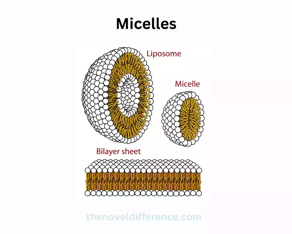
Size and structure of micelles
Here’s a simplified breakdown of the size and structure of micelles:
- Tiny Structures: Micelles are extremely small structures formed in aqueous solutions.
- Spherical Shape: They are typically spherical in shape, composed of amphiphilic molecules, such as bile salts or phospholipids, arranged with their hydrophobic tails clustered inward and hydrophilic heads facing outward.
- Nanometer Scale: Micelles are very small, with diameters ranging from about 5 to 100 nanometers, making them significantly smaller than cells.
- Core-Shell Structure: They have a core made up of hydrophobic (water-repellent) tails of the amphiphilic molecules, while the hydrophilic (water-attracting) heads form the outer shell, allowing them to solubilize and transport fat-soluble substances in aqueous environments.
- Dynamic Nature: Micelles are dynamic structures, continuously forming and breaking down, crucial for aiding in the digestion and absorption of dietary fats by facilitating the transport of lipids through the watery environment of the intestines.
Micelles are tiny, spherical structures composed of amphiphilic molecules arranged in a manner that enables the solubilization and transport of fats and fat-soluble substances in aqueous environments like the intestines during the process of digestion and absorption.
What are Chylomicrons?
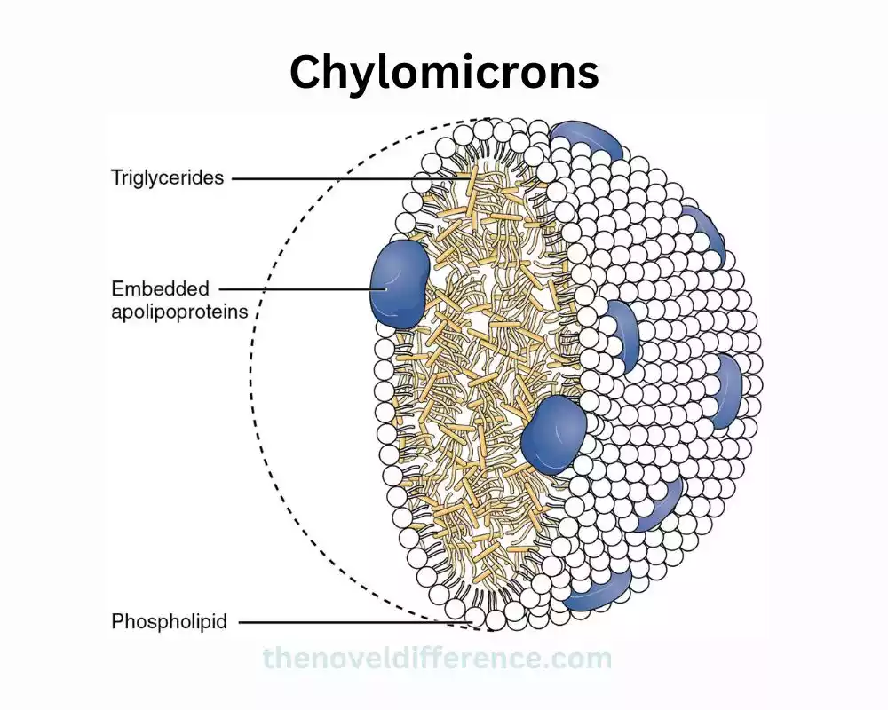
Chylomicrons are large, spherical particles essential for transporting dietary fats throughout the body. They are primarily formed in the small intestine after the absorption of dietary lipids, such as triglycerides, cholesterol, and fat-soluble vitamins.
After consuming a meal rich in fats, the small intestine absorbs these dietary fats. Inside the intestinal cells, these fats are reassembled into triglycerides. These triglycerides are then combined with proteins and phospholipids to form chylomicrons, creating these specialized particles.
Once formed, chylomicrons are released into the lymphatic system and then into the bloodstream, where they act as vehicles for transporting fats and fat-soluble vitamins to various tissues and organs throughout the body. They’re particularly crucial for delivering dietary lipids to tissues like adipose (fat) tissue, muscle cells, and the liver.
Chylomicrons are relatively large in size compared to other lipoproteins and are primarily composed of triglycerides in their core, surrounded by a shell consisting of proteins, phospholipids, and cholesterol. As they travel through the bloodstream, chylomicrons gradually release their triglyceride content to cells requiring energy or storage.
Eventually, chylomicrons undergo enzymatic breakdown by lipoprotein lipase, releasing fatty acids for energy or storage, and the remnants are taken up by the liver for further processing or excretion. In essence, chylomicrons play a crucial role in the absorption and transport of dietary fats, supporting various bodily functions and energy needs.
Formation of chylomicrons
Here’s a simplified breakdown of the formation of chylomicrons:
- Dietary Fat Absorption: When you consume dietary fats (triglycerides) through food, they travel to the small intestine for absorption.
- Reassembly in Intestinal Cells: Inside the cells lining the small intestine, these dietary fats are reassembled into triglycerides.
- Combination with Proteins: The reassembled triglycerides are combined with proteins (apoproteins), cholesterol, and phospholipids within these intestinal cells.
- Formation of Chylomicrons: The amalgamation of triglycerides, proteins, cholesterol, and phospholipids results in the creation of specialized particles called chylomicrons.
- Release into Lymphatic System: Chylomicrons are released into the lymphatic system through lacteals (lymphatic vessels in the intestines) and eventually enter the bloodstream.
- Transport of Dietary Fats: Once in the bloodstream, chylomicrons act as vehicles for transporting dietary fats, particularly triglycerides and fat-soluble vitamins, to various tissues and organs throughout the body.
Micelles and Chylomicrons in the comparison chart
| Aspect | Micelles | Chylomicrons |
| Size | Very small, typically ranging from 5 to 100 nanometers | Relatively large particles, several hundred nanometers in diameter |
| Formation Location | Formed in the intestines during lipid digestion | Formed in the small intestine after absorption of dietary fats |
| Composition | Composed of amphiphilic molecules (e.g., bile salts) | Comprised of triglycerides, proteins, cholesterol, and phospholipids |
| Function | Solubilize and transport fat-soluble substances in aqueous environments | Transport dietary fats, particularly triglycerides and fat-soluble vitamins, throughout the body |
| Transported Substances | Facilitate absorption of fat-soluble vitamins and fatty acids | Carry dietary lipids to various tissues and organs |
| Role in Digestion | Aid in the digestion and absorption of dietary fats | Contribute to the transport and utilization of dietary fats for energy or storage |
| Structure | Spherical structures with a core-shell configuration | Large spherical particles with a core of triglycerides and a surface layer of proteins, cholesterol, and phospholipids |
| Metabolic Fate | Break down after lipid absorption, reform during digestion | Broken down by lipoprotein lipase, releasing fatty acids for energy or storage, and remnants are taken up by the liver |
Micelles and chylomicrons both play critical roles in lipid metabolism, but they differ significantly in size, composition, function, and the types of lipids they transport and solubilize within the body. Understanding their differences is essential in comprehending the digestion, absorption, and transport of dietary fats.
Similarities of Micelles and Chylomicrons
Here are the similarities between micelles and chylomicrons:
- Lipid Transport: Both micelles and chylomicrons are involved in transporting lipids (fats) within the body.
- Formation in the Intestines: They are formed in the intestines during the process of lipid digestion and absorption.
- Emulsification of Fats: Both structures aid in the solubilization and transport of dietary lipids, making them available for absorption in the intestines.
- Transport of Fat-Soluble Substances: Both micelles and chylomicrons assist in transporting fat-soluble vitamins (such as vitamins A, D, E, and K) and fatty acids.
- Size Variation: Both structures vary in size – micelles are small, while chylomicrons are relatively larger, aiding in their specific roles in lipid transport within the body.
While micelles and chylomicrons share similarities in their involvement in lipid transport and the absorption of fat-soluble substances, they differ in their specific structures, functions, and the types of lipids they transport.
Conclusion
Micelles and chylomicrons are distinct entities involved in the transportation of fats within the body. Micelles aid in fat absorption during digestion, while chylomicrons transport dietary fats to various tissues. Understanding the Differences Between Micelles and Chylomicrons is Essential for Comprehending the intricate Mechanisms That Ensure the Efficient Transport and Utilization of Fats in the Human Body.