 by
by Understanding the complex workings of the human body may be a captivating endeavor. The field of life systems and physiology is wealthy with complex structures and components that contribute to our capacity to move and perform different assignments. Two essential components in the muscular system are the muscle spindle and the Golgi tendon organ. These sensory receptors play distinct roles in regulating muscle function. In this comprehensive guide, we will explore the difference between the muscle spindle and Golgi tendon organ, shedding light on their unique characteristics, functions, and contributions to our overall movement and coordination.
Importance of sensory receptors in muscle function
Sensory receptors play a vital role in the complex functioning of muscles. These receptors provide essential information about the state of muscles, enabling our bodies to maintain balance, coordination, and control over movement. Among the different tactile receptors within the musculoskeletal framework, two especially imperative ones are the muscle axle and the Golgi ligament organ.
The muscle spindle is responsible for monitoring muscle length and stretch. It is located within the muscle belly and consists of specialized intrafusal muscle fibers. When a muscle is extended, the muscle axle is actuated, and tangible neurons pass on this data to the central anxious framework. This feedback allows the brain to adjust muscle tone, initiate appropriate muscle contractions, and generate reflexes. The muscle spindle’s part in muscle coordination and proprioception (mindfulness of body position) is pivotal for keeping appropriate poses, executing exact developments, and adjusting to environmental changes.
The Golgi tendon organ is positioned within the tendon, where it detects muscle tension and force generation. It consists of encapsulated sensory receptors that respond to increased tension in the tendon. Sensory neurons transmit this information to the central nervous system. The Golgi tendon organ acts as a protective mechanism, preventing excessive muscle force that could potentially lead to injury. It also contributes to fine motor control by regulating muscle contraction and force production, enabling delicate and precise movements.
The sensory feedback provided by these receptors is essential for our body’s ability to execute movements effectively and safely. By constantly monitoring muscle length, stretch, tension, and force, the sensory receptors contribute to the overall coordination, balance, and stability of our musculoskeletal system. They assist in adjusting muscle activity, maintaining appropriate muscle tone, and preventing potential damage due to excessive force. Without these sensory receptors, our ability to perform complex movements and maintain physical well-being would be significantly compromised.
Understanding the significance of tactile receptors in muscle work has significant suggestions for different areas such as sports science, restoration, and neurology. By studying and harnessing the information provided by the muscle spindle and Golgi tendon organ, researchers and healthcare professionals can develop strategies to optimize performance, enhance rehabilitation outcomes, and address motor disorders. An advanced investigation in this range opens the potential applications and benefits that can be inferred from a more profound understanding of these tangible receptors and their part in muscle work.
Definition of Spindle and Golgi Tendon Organ
Muscle Spindle: The muscle spindle is a sensory receptor located within the muscle belly. It is composed of specialized intrafusal muscle fibers that run parallel to the regular muscle fibers (extrafusal fibers). The muscle shaft is mindful of checking muscle length, changes in muscle length (extend), and the rate of alteration in muscle length. It gives vital proprioceptive data approximately the position and development of the muscles to the central apprehensive framework. When the muscle is extended, the muscle axle is enacted, driving the transmission of tangible signals to the brain and spinal rope, which in turn start suitable muscle reactions, such as muscle compression and reflexes.
Golgi Tendon Organ: The Golgi tendon organ (GTO) is another sensory receptor located within the tendons, near their junction with the muscles. It consists of specialized encapsulated nerve endings intertwined among the collagen fibers of the tendon. The GTO is responsible for monitoring muscle tension and force generation. It faculties change in muscle pressure that happens amid muscle withdrawal, and it gives critical criticism to the central anxious framework almost the sum of the drive being produced by the muscle. When the pressure within the ligament increments, the Golgi ligament organ is actuated, driving the transmission of tangible signals to the brain and spinal line.
The GTO’s primary function is to protect the muscle from excessive force and injury by causing muscle relaxation through reflex pathways when the tension becomes too high. The muscle spindle and Golgi’s tendon organ are sensory receptors that play crucial roles in providing feedback about muscle length, stretch, tension, and force to the central nervous system. They contribute to maintaining proper muscle tone, coordinating muscle activity, and protecting muscles from potential damage.
What is Muscle Spindle?
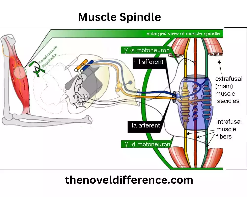
The muscle spindle is a specialized sensory receptor located within the muscle belly. It is composed of intrafusal muscle fibers, which are muscle fibers that are distinct from the regular muscle fibers (extrafusal fibers) responsible for generating force and movement. The muscle axle capacities as a proprioceptive organ, giving data approximately the length and extent of the muscle to the central anxious framework.
The structure of the muscle spindle includes sensory endings called primary and secondary endings, which are wrapped around the intrafusal muscle fibers. These tangible endings are associated with tactile neurons that transmit signals to the spinal line and brain. Engine neurons innervate the intrafusal muscle filaments, permitting them to contract and alter the affectability of the shaft.
When a muscle is stretched, the muscle spindle is activated. The intrafusal muscle fibers within the spindle are elongated, and this elongation causes deformation of the sensory endings. This deformation generates action potentials in the sensory neurons, which transmit signals to the spinal cord and brain. These signals provide important information about the length, rate of change, and direction of muscle stretch.
The sensory feedback from the muscle spindle is crucial for maintaining proper muscle tone, coordinating muscle activity, and enabling precise movements. It helps in detecting changes in muscle length, whether caused by external forces or voluntary contractions and allows for immediate adjustments in muscle contraction and reflex responses. This feedback loop between the muscle spindle, sensory neurons, and the central nervous system ensures accurate control of muscle movement and posture.
The muscle axle may be a specialized tactile receptor that plays an essential part in proprioception, giving basic data about muscle length and extending to the central anxious framework. It contributes to the coordination, control, and fine-tuning of muscle activity, allowing for efficient movement and motor function.
Structure and location
The muscle spindle is a specialized structure located within the muscle belly. It is composed of intrafusal muscle fibers, which are distinct from the regular muscle fibers (extrafusal fibers) responsible for generating force and movement.
The structure of the muscle spindle consists of several key components:
1. Intrafusal Muscle Fibers: These are muscle fibers that make up the bulk of the muscle spindle. Unlike the extrafusal fibers responsible for muscle contraction and force production, intrafusal fibers are smaller and specialized for sensory functions rather than generating force. They are encapsulated within a connective tissue sheath and run parallel to the extrafusal muscle fibers.
2. Sensory Endings: The intrafusal muscle fibers are surrounded by sensory endings, also known as sensory receptors or sensory corpuscles. These sensory endings are of two types:
a. Primary Endings (Annulospiral Endings): Primary endings are larger, more numerous, and wrap around the central region of the intrafusal muscle fibers. They are highly sensitive to changes in muscle length and velocity and provide accurate information about muscle stretch.
b. Secondary Endings (Flower Spray Endings): Secondary endings are smaller in size and are located near the ends of the intrafusal muscle fibers. They are less sensitive to changes in muscle length and velocity than primary endings.
3. Motor Innervation: The muscle spindle receives motor innervation from gamma motor neurons. These gamma motor neurons innervate the ends of the intrafusal muscle fibers, allowing for their contraction. By adjusting the sensitivity and tension of the intrafusal fibers, the gamma motor neurons modulate the responsiveness and effectiveness of the muscle spindle.
The muscle spindle is distributed throughout the muscle, with multiple spindles present within a single muscle belly. They are most abundant in muscles responsible for fine movements and precise control, such as those found in the hand and fingers. The exact number and arrangement of muscle spindles vary depending on the specific muscle and its functional requirements.
The muscle spindle’s structure, composed of intrafusal muscle strands, tactile endings, and engine innervation, is particularly outlined to identify changes in muscle length and give proprioceptive criticism to the central anxious framework, contributing to the coordination and control of muscle action.
Function
The muscle spindle serves several important functions related to sensory feedback and muscle control.
These functions include:
1. Sensory Role in Muscle Length and Stretch: The primary function of the muscle spindle is to monitor muscle length and changes in muscle length (stretch). When a muscle is stretched, the muscle spindle is activated, and sensory receptors within the spindle detect the extent and rate of stretch. This information is then transmitted to the central nervous system, providing crucial feedback about the position and movement of the muscle.
2. Contribution to Muscle Contraction and Reflexes: The muscle spindle plays a significant role in muscle contraction and reflexes. When the muscle spindle detects a stretch, sensory neurons associated with the spindle transmit signals to the spinal cord and brain. This sensory input is integrated with other sensory information, allowing for appropriate adjustments in muscle contraction. The muscle spindle contributes to the regulation of muscle tone and the coordination of muscle activity, ensuring precise movement and control.
3. Proprioception and Body Awareness: Proprioception refers to the ability to sense and perceive the position, movement, and orientation of one’s body parts. The muscle spindle is a critical component of proprioceptive feedback, providing information about muscle length and changes in muscle length. This feedback is essential for maintaining body awareness, balance, and posture. It enables individuals to accurately perceive the position of their limbs and control their movements without relying solely on visual cues.
4. Contribution to Motor Learning and Coordination: The sensory feedback provided by the muscle spindle is crucial for motor learning and coordination. It permits people to fine-tune their developments and make exact alterations based on the tangible data gotten from the muscle shaft. Through repeated sensory input and integration with other sensory modalities, the muscle spindle contributes to the development of skilled movements and the refinement of motor control.
5. Detection of Muscle Abnormalities or Imbalances: The muscle spindle is sensitive to changes in muscle length and tension. This sensitivity enables it to detect abnormalities or imbalances in muscle function. If there’s a muscle shortcoming or an issue with muscle coordination, the muscle axle can give input that can offer assistance in recognizing and addressing these issues. By detecting and signaling such issues, the muscle spindle plays a role in maintaining muscle health and optimizing overall muscle function.
The muscle axle serves as a specialized tactile receptor that plays a vital part in giving input around muscle length, extension, and changes in muscle position to the central anxious framework. It contributes to muscle contraction, reflexes, proprioception, motor learning, and coordination. By continuously monitoring muscle length and providing real-time sensory information, the muscle spindle ensures accurate control of muscle activity and enables individuals to perform coordinated and precise movements.
What is the Golgi Tendon Organ?
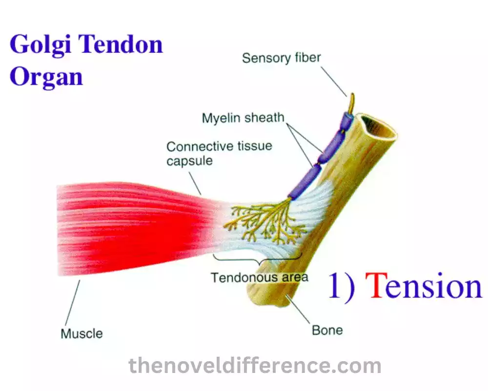
The Golgi tendon organ (GTO) is a sensory receptor located within the tendons, near their junction with the muscles. It is named after Italian physician Camillo Golgi, who first described it. The GTO functions as a proprioceptive organ, providing information about muscle tension and force generation to the central nervous system.
The structure of the Golgi tendon organ consists of several components:
1. Sensory Endings: The GTO contains specialized encapsulated nerve endings known as Golgi tendon receptors. These receptors are intertwined among the collagen fibers of the tendon. The sensory endings are highly sensitive to changes in tendon tension and are connected to sensory neurons.
2. Connective Tissue Capsule: The sensory endings of the GTO are encapsulated within a connective tissue capsule, providing protection and support.
3. Junction with Muscle Fibers: The GTO is situated at the junction of muscle fibers and the tendon, allowing it to sense the tension produced during muscle contraction and the resulting force exerted on the tendon.
When tension is applied to the tendon, such as during muscle contraction or external resistance, the Golgi tendon organ is activated. The tension causes deformation of the collagen fibers within the tendon, which, in turn, stimulates the sensory receptors of the GTO. This stimulation generates action potentials in the sensory neurons connected to the GTO.
The sensory signals from the Golgi tendon organ are then transmitted to the central nervous system, specifically to the spinal cord and brain. This feedback provides essential information about the amount of force being generated by the muscle. It allows the central nervous system to regulate muscle activity and maintain appropriate force production based on the body’s needs.
One of the primary functions of the Golgi tendon organ is to protect the muscle from excessive force and prevent injury. When the tension on the tendon becomes too high, the GTO reflexively triggers a relaxation response in the muscle. This defensive instrument prevents the muscle from applying as well as much constrain, which might harm the muscle or its connections.
The Golgi tendon organ also contributes to fine motor control and precision. Providing feedback about the level of tension in the muscle-tendon unit helps regulate the force output during delicate movements. This allows for precise control and coordination of muscle activity, especially in tasks that require intricate motor skills.
The Golgi tendon organ is a specialized sensory receptor located at the junction of muscle fibers and tendons. It detects changes in tendon tension and provides sensory feedback to the central nervous system. The GTO plays a crucial role in protecting muscles from excessive force, contributing to motor control, and enabling precise movements.
Clinical significance
The clinical significance of the Golgi tendon organ (GTO) lies in its role as a diagnostic tool and its impact on rehabilitation and injury prevention.
Here are some clinical considerations related to the GTO:
1. Diagnostic Tool: The GTO can be utilized in the clinical setting to assess muscle function and detect abnormalities. By assessing the response of the GTO during manual muscle testing or specific joint movements, healthcare professionals can gather valuable information about muscle strength, coordination, and neuromuscular control. Abnormal responses, such as an overactive GTO leading to excessive inhibition of muscle contraction, can provide insights into muscle imbalances, weakness, or neurological disorders.
2. Injury Prevention and Rehabilitation: The GTO plays a crucial role in injury prevention and rehabilitation. Its protective reflex mechanism prevents excessive force generation in the muscle-tendon unit, which can help reduce the risk of muscle strains, tendon injuries, and muscle imbalances. Understanding the function of the GTO can guide the development of rehabilitation programs and exercise protocols to optimize muscle strength, flexibility, and neuromuscular control while ensuring proper load management to avoid overloading the tissues.
3. Neuromuscular Control and Motor Learning: The sensory feedback provided by the GTO contributes to neuromuscular control and motor learning. By integrating information about muscle tension, the central nervous system can adjust muscle activation patterns, muscle recruitment, and coordination to optimize movement execution. Rehabilitation programs can incorporate exercises that target GTO activation and sensory feedback to enhance motor control, proprioception, and kinesthetic awareness.
4. Performance Optimization: Understanding the role of the GTO in regulating force production can be beneficial for athletes and individuals involved in sports and physical activities. By targeting specific training techniques and exercises that stimulate the GTO, it is possible to enhance neuromuscular control, optimize force generation, and improve performance. This information can be connected to sports-specific preparation, quality, and conditioning programs, and harm avoidance procedures.
5. Rehabilitation after Injury or Surgery: Following injury or surgical interventions, the GTO’s function and sensory feedback may be affected. Physical therapy and rehabilitation protocols can focus on retraining the GTO response, improving muscle-tendon unit coordination, and gradually reintroducing appropriate loads and forces to the injured tissues. This process helps restore normal muscle function, proprioception, and strength, promoting successful recovery and preventing re-injury.
The Golgi tendon organ has clinical significance in various areas of healthcare. It serves as a diagnostic tool, contributes to injury prevention and rehabilitation, influences neuromuscular control and motor learning, aids in performance optimization, and guides restoration after injury or surgery. Understanding and utilizing the clinical implications of the GTO can help improve patient outcomes, enhance athletic performance, and promote overall musculoskeletal health.
Function
The Golgi tendon organ (GTO) serves several important functions related to muscle function and control.
Here are the key functions of the GTO:
1. Monitoring Muscle Tension: The primary function of the GTO is to monitor muscle tension or force generated during muscle contraction. As muscles contract, the tension produced is transmitted to the tendons. The GTO detects these changes in tendon tension and provides sensory feedback about the level of force being exerted by the muscle.
2. Regulation of Muscle Contraction: The sensory information provided by the GTO plays a role in regulating muscle contraction. When the GTO recognizes intemperate pressure or drives within the muscle, it starts a defensive reflex called the “Golgi ligament reflex.” This reflex leads to the restraint of muscle withdrawal, causing the muscle to unwind. This mechanism helps prevent the muscle from generating excessive force that could potentially damage the muscle or its attachments.
3. Load Distribution and Force Control: The GTO contributes to load distribution and force control within a muscle-tendon unit. Monitoring tension at the muscle-tendon junction helps distribute the force evenly across the muscle fibers and tendons. This ensures efficient force transmission and minimizes the risk of localized overload or strain on specific muscle fibers or tendons.
4. Proprioception and Joint Stability: The sensory feedback from the GTO plays a role in proprioception, which refers to the sense of body position and movement. The GTO provides information about muscle tension, which contributes to proprioceptive awareness and joint position sense. This information helps in maintaining joint stability and coordinating muscle activity during various movements and tasks.
5. Coordinated Muscle Control: The GTO, along with other sensory receptors and neural pathways, contributes to coordinated muscle control. By providing feedback about muscle tension, the GTO helps in coordinating muscle activity, regulating muscle synergies, and adjusting muscle recruitment patterns. This coordination is crucial for smooth and efficient movement, as well as for maintaining postural stability.
6. Force Regulation and Injury Prevention: The GTO acts as a protective mechanism to prevent excessive force generation in the muscle-tendon unit. By triggering the Golgi tendon reflex, it inhibits muscle contraction when tension becomes too high, thus protecting the muscle and its attachments from potential damage. This function helps prevent muscle strains, tendon injuries, and overload-related conditions.
The Golgi tendon organ plays a vital role in monitoring muscle tension, regulating muscle contraction, contributing to proprioception and joint stability, facilitating coordinated muscle control, and preventing excessive force generation. By providing sensory feedback about muscle tension, the GTO helps maintain optimal muscle function, protect against injuries, and ensure efficient force control during movement and physical activities.
Comparison Chart
Sure! Here’s a comparison chart highlighting the key differences between the muscle spindle and Golgi tendon organ (GTO):
| Muscle Spindle | Golgi Tendon Organ (GTO) |
|---|---|
| Located within the muscle belly, embedded among muscle fibers. | Located at the junction of the muscle and tendon, within the tendon itself. |
| Consists of intrafusal muscle fibers and sensory endings that wrap around these fibers. | Consists of specialized sensory endings within a connective tissue capsule. |
| Detects changes in muscle length and the rate of change of muscle length. Provides feedback on muscle stretch. Involved in proprioception, muscle contraction, reflexes, and coordination. | Detects changes in muscle tension or force generated during muscle contraction. Provides feedback on the amount of force exerted by the muscle on the tendon. Contributes to force regulation, load distribution, and prevention of excessive force generation. |
| Contains primary and secondary sensory endings that wrap around intrafusal muscle fibers. | Contains Golgi tendon receptors intertwined among the collagen fibers of the tendon. |
| Can elicit the stretch reflex, leading to the contraction of the muscle in response to its stretch. | Can elicit the Golgi tendon reflex, leading to the relaxation and inhibition of the muscle in response to excessive tension. |
| Provides information about muscle length, stretch, and muscle velocity to the central nervous system. | Provides information about tendon tension or force being exerted by the muscle on the central nervous system. |
| Not a direct protective mechanism, but contributes to muscle tone and stability. | Acts as a protective mechanism by inhibiting excessive force generation in the muscle-tendon unit. |
| Plays a crucial role in proprioception by providing feedback on muscle position, movement, and muscle fiber length. | Contributes to proprioception by providing feedback on force production and load distribution within the muscle-tendon unit. |
This comparison chart summarizes the main differences between the muscle spindle and Golgi tendon organ in terms of their location, structure, function, sensory input, reflex responses, and role in proprioception and protective mechanisms.
Conclusion
The muscle spindle and Golgi tendon organ are distinct sensory receptors that provide essential feedback about muscle length and tension. While the muscle spindle monitors muscle length and initiates reflex responses for precise muscle control, the Golgi tendon organ acts as a protective mechanism against excessive tension.
Understanding the difference between these sensory receptors deepens our knowledge of how our bodies move and function. By appreciating their roles, we can enhance our performance, prevent injuries, and maintain a healthy musculoskeletal system.